. 1996 Nov 1;16(21):6839-52.
doi: 10.1523/JNEUROSCI.16-21-06839.1996.

Characterization of densin-180, a new brain-specific synaptic protein of the O-sialoglycoprotein family

Affiliations

Characterization of densin-180, a new brain-specific synaptic protein of the O-sialoglycoprotein family

M L Apperson et al. J Neurosci. .

Abstract

We purified an abundant protein of apparent molecular mass 180 kDa from the postsynaptic density fraction of rat forebrain and obtained amino acid sequences of three tryptic peptides generated from the protein. The sequences were used to design a strategy for cloning the cDNA encoding the protein by polymerase chain reaction. The open reading frame of the cDNA encodes a novel protein of predicted molecular mass 167 kDa. We have named the protein densin-180. Antibodies raised against the predicted amino and carboxyl sequences of densin-180 recognize a 180 kDa band on immunoblots that is enriched in the postsynaptic density fraction. Immunocytochemical localization of densin-180 in dissociated hippocampal neuronal cultures shows that the protein is highly concentrated at synapses along dendrites. The message encoding densin-180 is brain specific and is more abundant in forebrain than in cerebellum. The sequence of densin-180 contains 17 leucine-rich repeats, a sialomucin domain, an apparent transmembrane domain, and a PDZ domain. This arrangement of domains is similar to that of several adhesion molecules, in particular GPIbalpha, which mediates binding of platelets to von Willebrand factor. We propose that densin-180 participates in specific adhesion between presynaptic and postsynaptic membranes at glutamatergic synapses.


Figures

Fig. 1.
Fig. 1.
Tryptic peptide sequences and PCR cloning of densin-180. A, Amino acid sequences of three tryptic peptides from densin-180 were used to design sense (S) and antisense (A) degenerate 29 mer oligonucleotide primers: A, adenosine; C, cytosine;G, guanosine; I, inosine;T, thymidine. Degenerate nucleotide positions are enclosed in parentheses. The 10 amino acids used to design the sense (right-pointing arrows) and antisense (left-pointing arrows) are indicated above and below the peptide sequences, respectively. B, PCR cloning strategy. Combinations of sense and antisense primers (arrows) were used for amplification of sequences from rat forebrain cDNA by PCR. A PCR product (represented by hatched box) was cloned into a vector, and its nucleotide sequence was determined by dideoxy sequencing from the M13 and T7 primer sites of the vector (bold arrows). C, DNA from PCR reactions was fractionated on a 1.2% agarose gel, and the DNA was visualized by ethidium bromide staining. The size of the PCR product was estimated by comparison with DNA molecular weight markers. The 1S + 3A primer combination produced a 1.2 kb PCR product (arrow) that was absent in reactions containing 1S or 3A alone. D, Restriction map and sequencing strategy for cDNA clones encoding densin-180. Clone 1.1 was sequenced in its entirety, and the coding region (open box), the region of hybridization with the PCR product (hatched box), and the 5′ and 3′ noncoding regions (horizontal lines) are indicated on the restriction map (map units are in base pairs). The locations of cDNA clones determined by restriction mapping and sequencing are shownbelow the restriction map. The extent and directionality of overlapping cDNA sequences are depicted as arrows for each cDNA clone. Clone 2.1 lacks nucleotides 111–186 of the densin-180 sequence (broken line) containing the ribosome-binding site and part of the initiation codon. Clone 3.1 lacks a 249 base pair sequence spanning nucleotides 1632–1880 (broken line) encoding amino acids 483–565 of the densin-180 sequence.
Fig. 2.
Fig. 2.
Protein sequence translated from the densin-180 cDNA. The DNA sequence of clone 1.1, containing the entire coding region, was determined by sequencing both strands. It has been deposited in the GenBank database and assigned accession number U66707. The protein translation is shown in the figure. Protein sequences of tryptic peptides 1, 2, and 3 are underlined. Potential N-linked glycosylation sites, CaMKII phosphorylation sites (bold), and RGD cell attachment motif are shown asboxed residues. The potential transmembrane domain isunderlined (gray bar), and the 16 leucine-rich repeats are contained in amino acids 53–420. The amino- and carboxy-flanking cysteine-rich domains span amino acids 19–37 and 486–546, respectively. The mucin homology domain spans amino acids 825–915, and the PDZ domain spans amino acids 1405–1492.
Fig. 3.
Fig. 3.
Domain structure of densin-180. A, Alignment of the 16 densin-180 leucine-rich repeats reveals a 23 residue consensus shown at the bottom. Corresponding amino acid numbers of densin-180 are indicated to theleft of the first repeat and to the rightof the 16th repeat. B, Identification of a PDZ domain in densin-180. Alignment of amino acids 1400–1493 of densin-180 with 10 PDZ domains from four other proteins [3 PDZ domains fromPSD95 (Cho et al., 1992); 3 fromDrosophila disks-large protein (DLG;Woods and Bryant, 1991); 3 from the human zona occludens protein (ZO-1; Itoh et al., 1993); 1 from neuronal nitric oxide synthase (nNOS; Bredt et al., 1991)]. C, The domain structure of densin-180 as compared with the LRR-containing glycoprotein GPIbα. The leucine-rich repeats (16 in densin-180 and 7 in GPIbα; wavy lines) with N-terminal and C-terminal cysteine-rich flanking regions (lightly stippled) and mucin-like domains (diagonal lines) are indicated for both proteins. Potential transmembrane domains are depicted in black. The ABP-binding protein domain of GPIbα and the PDZ domain of densin-180 at the C-terminal regions of the proteins are represented ingray. The position of the RGD sequence is indicated with an arrowhead. Scale bar, 150 amino acids.
Fig. 4.
Fig. 4.
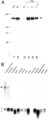
Densin-180 protein is enriched in PSD fractions, and its mRNA expression is brain-specific. A, Enrichment of densin-180 protein in detergent-extracted PSD fractions. Immunoblots were prepared with 50 μg (lanes 1, 2) of rat brain homogenate (Hom) and synaptosome fractions (Syn) and 7.5 μg (lanes 3–6) each of synaptosome (Syn), once Triton X-100-extracted PSD (1T), twice Triton X-100-extracted PSD (2T), and once Triton X-100 and then sarcosyl-extracted PSD (1T + S; Cho et al., 1992). Densin-180 protein band (arrow) is visualized with antibody M2 against densin-180. Molecular weight markers and position of the dye front (open arrowhead) are shown at left. B, Densin-180 Northern blot. Poly(A)+ RNA (5 μg) from 13 different tissue samples was electrophoresed on a 1% agarose gel. The mRNA was transferred to Zeta-Probe blotting membrane (Bio-Rad), and all lanes were determined to have equal amounts of RNA by methylene blue staining. Blots were probed with a random prime-labeled PCR-amplified DNA fragment of densin-180 spanning nucleotides 1100–2170 (specific activity, 109 cpm/μg). A single broad band at 7.4 kb was detected (large arrow) on autoradiographs exposed for 14 d with an intensification screen. The blot was then stripped and reprobed with the 2 kb random prime-labeled human β-actin cDNA (specific activity, 107 cpm/μg). The autoradiograph of an 8 hr exposure with an intensification screen is shown in the bottom panel. The two forms of β-actin message are indicated (small arrows).
Fig. 5.
Fig. 5.
Densin-180 is a mucin-like sialoglycoprotein.A, Densin-180 is heavily glycosylated with sialic acid. Twenty micrograms of denatured protein from the PSD fraction were incubated overnight at 37°C under each of the following conditions: control reaction with no added neuraminidase (lane 1), with added neuraminidase (lane 2), and with added neuraminidase plus 10 mmN-bromosuccinimide (lane 3). Digested protein was fractionated by SDS-PAGE and probed with antibody against densin-180, as described under Materials and Methods. The 188 kDa undigested (top) and 148 kDa digested (bottom) densin-180 protein bands are indicated by arrows. The positions of 205, 118, and 87 kDa molecular weight markers are shown at left.B, C, Proteolysis of densin-180 byO-sialoglycoprotein endoprotease. Nondenatured PSD fraction (24 μg) was incubated with 0.4 mg/ml final volume ofO-sialoglycoprotein endoprotease, as described under Materials and Methods. All incubations were performed in the presence of 0.2 mm PMSF to inhibit endogenous proteases in the PSD fraction. Protease reactions were incubated at 37°C for 15 min (15m), 1 hr (1h) and 3 hr (3h). Control reactions with no protease added were incubated for 3 hr at 37°C. Reactions were terminated by adding gel sample buffer and boiling for 3 min. Digested protein was fractionated by SDS-PAGE and probed with two different antibodies against densin-180. The immunoblot shown in B was probed with CT245, a rabbit polyclonal serum that reacts with epitopes in the potential cytoplasmic domain spanning residues 1374–1495 of densin-180. The CT245 antibody detects the undigested 188 kDa densin-180 protein band (large black arrow) and a complex pattern of proteolytic fragments (gray arrows) of densin-180. These proteolytic fragments include major bands at ∼70, 45 (doublet), 40 (doublet), 30, and 20 kDa. The 20 kDa band (large gray arrow) is resistant to proteolysis after 3 hr at 37°C. The immunoblot shown inC was prepared with M2, a mouse polyclonal ascites that reacts with epitopes contained in amino acids 466–958 of the putative extracellular domain of densin-180. This antibody detects the undigested 188 kDa densin-180 protein band (large black arrow) and 140 and 120 kDa proteolytic fragments (gray arrows). The positions of molecular weight standards are shown at the right side with open arrowheads indicating the origin of the gel (top) and dye front (bottom).
Fig. 6.
Fig. 6.
Solubility of densin-180 in brain membrane fractions. Crude membrane fractions were isolated from rat brain. Pellet (P) and supernatant (S) fractions were separated by centrifugation at 170,000 × gafter extraction of membranes with 1 m NaCl, 2% CHAPS, 1 m NaCl + 2% CHAPS, 2% Triton X-100, 1 m NaCl + 2% Triton X-100, or 0.2 m sodium bicarbonate, pH 11, for 1 hr at 4°C. Proteins were fractionated by SDS-PAGE and probed with antibody against densin-180. The position of the densin-180 band is indicated with an arrow.
Fig. 7.
Fig. 7.
Densin-180 is phosphorylated by endogenous CaMKII in the PSD fraction. Phosphorylation reactions containing32P-labeled ATP and 24 μg of protein from the PSD fraction were performed in the absence (lane 1) and presence (lane 2) of calcium and in the presence of calcium plus inhibiting antibodies to CaMKII (lane 3), as described under Materials and Methods. Reactions were terminated by adding SDS to a final concentration of 1% and boiling for 5 min. Densin-180 was immunoprecipitated from the denatured phosphorylation reactions with M2 antibody to densin-180, as described under Materials and Methods, and applied to a 6% SDS- polyacrylamide gel. A section of the autoradiograph of a 16 hr exposure of the dried gel is shown. The position of densin-180 is indicated by an arrow.
Fig. 8.
Fig. 8.
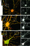
Immunocytochemical localization of densin-180 at synapses in dissociated hippocampal neurons. A–C, Hippocampal neurons dissociated at E18 were grown in culture on coverslips for 14–21 d and fixed with ice-cold methanol. After coverslips were incubated for 1 hr in preblock and overnight with the indicated pairs of primary antibodies, cultures were washed three times with preblock and incubated with Cy3-conjugated goat anti-mouse and FITC-conjugated goat anti-rabbit secondary antibodies. The coverslips then were washed and mounted on slides. Procedures are described in detail under Materials and Methods. Images were taken with a Zeiss laser-scanning fluorescence confocal microscope, and images of double-labeled cells were combined and colorized with Adobe Photoshop software. Red pseudocolor represents Cy3 staining, andgreen represents FITC staining. Regions of overlap areyellow. A, Double-staining for synapsin I and densin-180. Cultures grown for 21 d in vitrowere double-labeled with anti-synapsin I (1:1000; green) and anti-densin-180 (M2, 1:150; red). A combined image taken with a 63× objective is shown. The inset atleft is a 3× zoom of the area included in thewhite box. Note the overlap in staining for densin-180 (large arrowheads) and synapsin I (small arrows). At right are the single images of densin-180 (top) and synapsin I (bottom).B, Double-staining for PSD-95 and densin-180. Cultures grown for 17 d in vitro were double-labeled with anti-PSD-95 (affinity-pure, 1:100; green) and anti-densin-180 (M2, 1:150; red). A combined image taken with a 63× objective at Zoom 1.5 is shown. The axon initial segment stained for densin-180 is indicated with an arrow. Theinset at left is a 2× zoom of the area included in the white box. Note the precise colocalization of PSD-95 staining and densin-180 staining at spine-like structures along dendrites (large arrowheads). Atright are the single images of densin-180 (top) and PSD-95 (bottom).C, Double-staining for αCaMKII and densin-180. Cultures grown for 14 d in vitro were double-labeled with anti-αCaMKII (6G9, 1:500; green) and anti-densin-180 (CT245, 1:3000; red). A combined image taken with a 63× objective at Zoom 2 is shown. Theinset at left is a 2× zoom of the area included in the white box. Note examples of colocalization of αCaMKII staining and densin-180 staining at spine-like structures along dendrites (large arrowheads). At right are the single images of densin-180 (top) and αCaMKII (bottom).

References

    1. Abdullah KM, Udoh EA, Shewen PE, Mellors A. A neutral glycoprotease of Pasteurella haemolytica A1 specifically cleaves O-sialoglycoproteins. Infect Immun. 1992;60:56–62. - PMC - PubMed
    1. Aebersold RH, Leavitt J, Saavedra RA, Hood LE, Kent SBH. Internal amino acid sequence analysis of proteins separated by one- or two-dimensional gel electrophoresis after in situ protease digestion on nitrocellulose. Proc Natl Acad Sci USA. 1987;84:6970–6974. - PMC - PubMed
    1. Andrews RK, Fox JEB. Identification of a region in the cytoplasmic domain of the platelet membrane glycoprotein-IB–IX complex that binds to purified actin-binding protein. J Biol Chem. 1992;267:18605–18611. - PubMed
    1. Bahr BA, Lynch G. Purification of an Arg-Gly-Asp selective matrix receptor from brain synaptic plasma membranes. Biochem J. 1992;281:137–142. - PMC - PubMed
    1. Brenman JE, Chao DS, Gee SH, McGee AW, Craven SE, Santillano DR, Wu Z, Huang F, Xia H, Peters MF, Froehner SC, Bredt DS. Interaction of nitric oxide synthase with the postsynaptic density protein PSD-95 and α-1-syntrophin mediated by PDZ domains. Cell. 1996;84:757–767. - PubMed

Publication types

MeSH terms

Substances

Associated data

LinkOut - more resources