Comparative Study
. 1996 Nov 1;16(21):6896-907.
doi: 10.1523/JNEUROSCI.16-21-06896.1996.

Differential survival of Cajal-Retzius cells in organotypic cultures of hippocampus and neocortex

Affiliations
Comparative Study

Differential survival of Cajal-Retzius cells in organotypic cultures of hippocampus and neocortex

J A Del Río et al. J Neurosci. .

Abstract

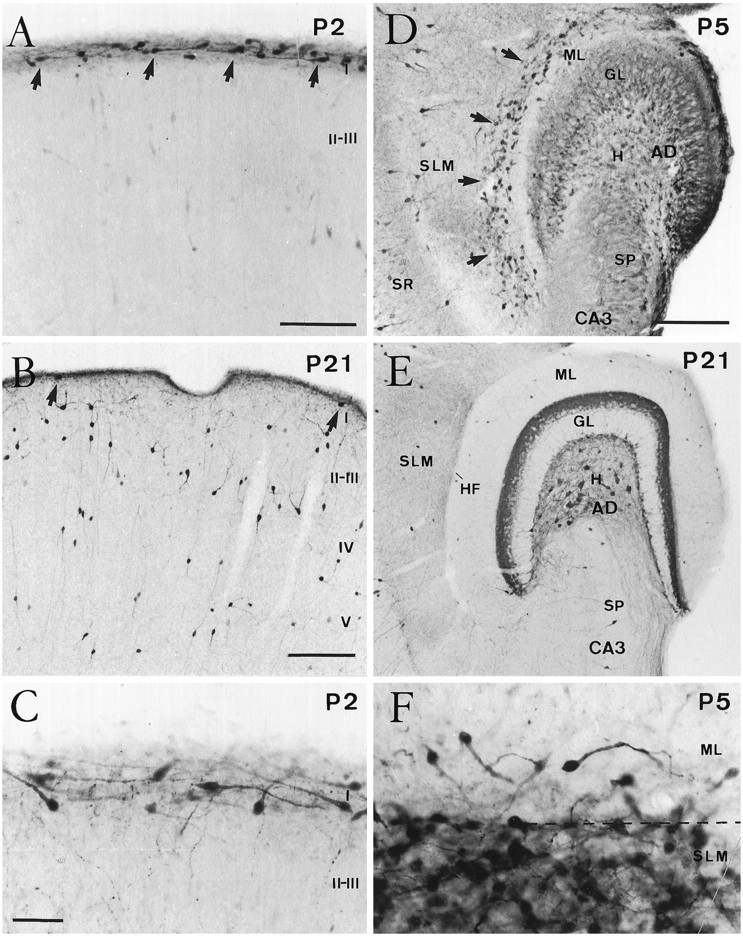
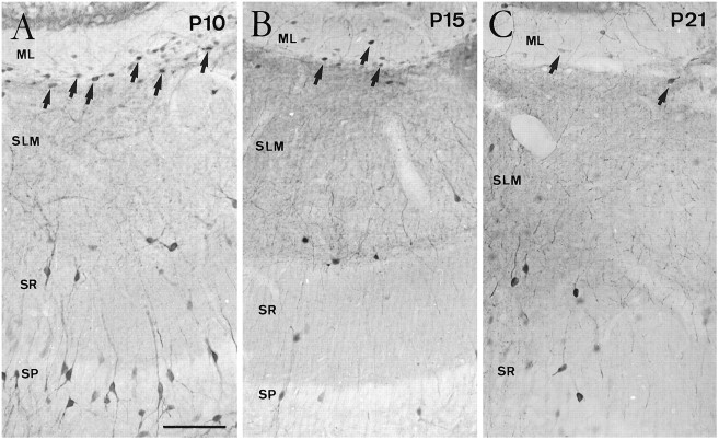
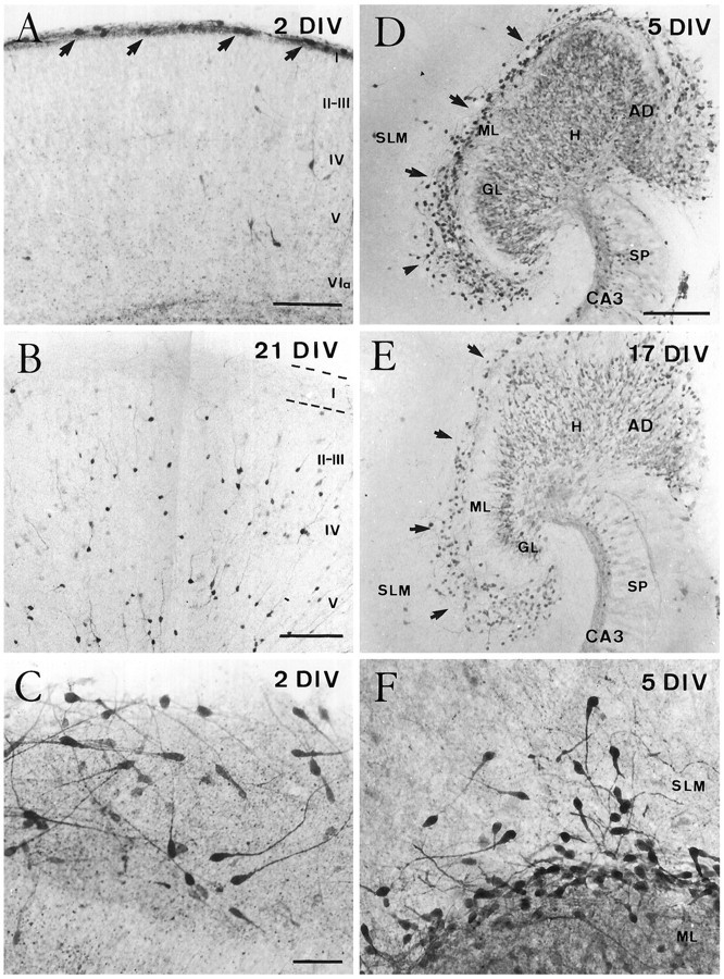
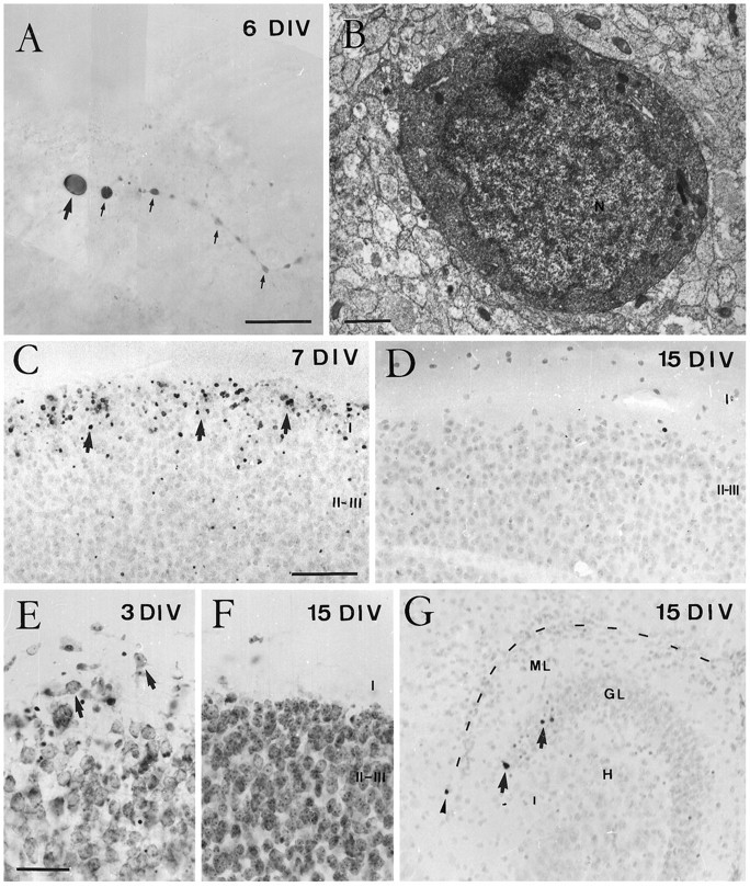
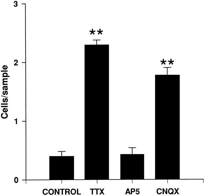
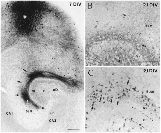
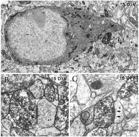
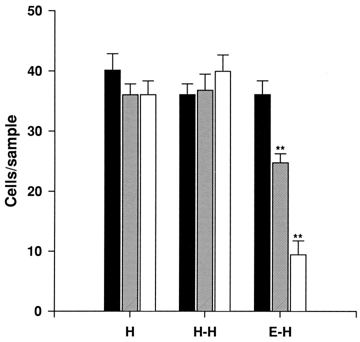
Cajal-Retzius (CR) cells are transient, pioneer neurons of layer I of the cortex that are believed to play essential roles in corticogenesis, e.g., in neuronal migration and synaptogenesis. Here we have used calretinin immunostaining to study the characteristics, survival, and fate of CR cells in single organotypic slice cultures of mouse neocortex and hippocampus deprived of their extrinsic afferents. In neocortical explants, CR cells were observed after 1-3 d in vitro (DIV), but they disappeared after 5-7 DIV, which is similar to their time of degeneration in vivo. The disappearance of CR cells in neocortical slices was prevented by incubation with tetrodotoxin and the glutamate receptor antagonist 6-cyano-7-nitroquinoxaline-2,3,-dione but not by 2-amino-5-phosphonopentanoic acid, suggesting that neuronal activity and non-NMDA glutamate receptors may trigger CR cell death in the neocortex. In contrast to the situation in vivo, in which many hippocampal CR cells disappear at approximately the third postnatal week, CR cells survived in single hippocampal cultures after long incubation times (31 DIV), with their morphology essentially unaltered. In contrast, fewer CR cells were found when hippocampal slices were cocultured with explants from the entorhinal cortex. Because CR cells are transient synaptic targets for entorhinohippocampal afferents, these findings suggest a role for entorhinal afferents in the degeneration of CR cells in the hippocampus. In conclusion, this study shows different survival properties of CR cells in organotypic slice cultures of hippocampus and neocortex, and it suggests that different mechanisms are involved in the regulation of the process of naturally occurring CR cell death in the two cortical regions.


Figures

Fig. 1.
Fig. 1.
Postnatal evolution of calretinin immunostaining in the developing neocortex and hippocampus. A, B, Coronal sections of the neocortex at P2 (A) and P21 (B). Although at P2 (A) numerous Cajal–Retzius cells (arrows) were present in layer I, at P21 (B) calretinin-positive cells in this layer were seen only rarely (arrows). In contrast, the number of calretinin-positive nonpyramidal neurons in layers II–V increased notably at P21. C, High-power photomicrograph showing the typical monopolar morphology of Cajal–Retzius cells in layer I at P2. D, E, Horizontal sections of the hippocampus at P5 (D) and P21 (E). At P5, Cajal–Retzius cells (arrows) are abundant near the hippocampal fissure, in the outer stratum lacunosum-moleculare (SLM), and in the adjacent aspect of the dentate molecular layer (ML). Immature granule cells in the granule cell layer (GL) displayed weak calretinin immunolabeling. At P21 (E), very few calretinin-positive neurons could be seen near the hippocampal fissure (HF). Detail shows densely packed Cajal–Retzius cells around the hippocampal fissure (dashed line) at P5. I–V, Cortical layers I–V; SR, stratum radiatum; H, hilus; SP, stratum pyramidale; CA3, hippocampal region CA3;AD, area dentata. Scale bars: A, 100 μm; B, 150 μm, also pertains to E;C, 50 μm, also pertains to F;D, 150 μm.
Fig. 2.
Fig. 2.
Evolution of Cajal–Retzius cells identified by calretinin immunolabeling in the late postnatal hippocampus. Although CR cells (arrows) are still abundant at P10 (A) in the stratum lacunosum-moleculare (SLM) and outer molecular layer (ML), substantial numbers of cells are observed at P15 (B) and fewer CR cells are present at P21 (C). Abbreviations as in Figure 1. Scale bar (shown in A): 100 μm, also pertains toB and C.
Fig. 3.
Fig. 3.
Evolution of Cajal–Retzius cells in organotypic slice cultures of the neocortex (A–C) and hippocampus (D–F) as shown by calretinin immunostaining. A, B, Cajal–Retzius cells (arrows) were seen in layer I in organotypic slices cultured for 2 DIV (A) but were virtually absent after long incubation times (B). Notice the increased immunolabeling of nonpyramidal neurons at 21 DIV. C, High magnification illustrating the morphological features of Cajal–Retzius cells after 2 DIV. D, E, Calretinin immunostaining in single organotypic slice cultures of hippocampus after 5 (D) and 17 (E) DIV. After both incubation times, Cajal–Retzius cells (arrows) were abundant in the molecular layer of the fascia dentata and in the stratum lacunosum-moleculare of the hippocampus proper. Weak immunolabeling was also seen in the granule cells and in the mossy fiber zone in CA3 region. F, High-magnification photomicrograph showing Cajal–Retzius cells in the stratum lacunosum-moleculare and in the dentate molecular layer of the hippocampus after 5 DIV. Abbreviations as in Figure 1. Scale bars:A, 150 μm; B, 200 μm;C, 50 μm, also pertains to F;D, 200 μm, also pertains to E.
Fig. 4.
Fig. 4.
Degeneration of Cajal–Retzius cells in organotypic slice cultures of neocortex. A, B, Correlated light (A) and electron microscopy (B) of a presumedly degenerating Cajal–Retzius cell after 6 DIV. A, At the light microscope, a calretinin-positive Cajal–Retzius cell displays shrunken cell body (arrow) and a swollen dendrite (small arrows). B, Electron micrograph of the perikaryon of the cell in A, showing disrupted fine structure of cytoplasmic organelles and chromatin condensation in the nucleus (N). C, D, Distribution of TUNEL-positive cells. In neocortical cultures, most TUNEL-positive cells (arrows) are present in layer I after 7 DIV (C), whereas TUNEL-positive cells are absent from this layer at 15 DIV (D). E, F, Distribution of Nissl-stained cell bodies in layer I of organotypic neocortical cultures. At short incubation times (E), some neurons in layer I display large perikarya and shapes typical of CR cells (arrows), whereas at longer incubation times (F) these large perikarya have disappeared from layer I. As in vivo, some pyknotic cells could also be noted at 3 DIV in layers II and III (E).G, Distribution of TUNEL-positive cells in hippocampal cultures. These cells are found in the stratum lacunosum-moleculare only very exceptionally, whereas some labeled neurons were observed in the granule cell layer (arrows). C, D,and G are from sections counterstained with hematoxylin. Abbreviations as in Figure 1. Scale bars: A, 25 μm;B, 1 μm; C, 100 μm, also pertains toD and G; E, 50 μm, also pertains to F.
Fig. 5.
Fig. 5.
The number of Cajal–Retzius cells present in layer I (number of cells per 250 μm horizontal stripe) in organotypic slice cultures of neocortex cultured for 10 DIV, after chronic treatment with TTX, AP5, or CNQX, as well as in control slices (mean ± SEM). Significant differences (**p ≤ 0.01; ANOVA, Scheffe’s test) compared with controls were found after TTX and CNQX treatments.
Fig. 7.
Fig. 7.
Reduction of hippocampal Cajal–Retzius cells in entorhinohippocampal cocultures. A, Low-power photomicrograph illustrating a typical entorhinohippocampal coculture after 7 DIV, in which entorhinal afferents to the hippocampus have been traced with biocytin. Section counterstained with hematoxylin. The injection site in the entorhinal cortex (EC) is marked by an asterisk. Anterogradely labeled entorhinal fibers (arrows) were seen in the stratum lacunosum-moleculare and in the dentate molecular layer. B, C, Distribution of Cajal–Retzius cells in an entorhinohippocampal coculture (B) and in a single hippocampal culture (C) after 21 DIV. A marked reduction in the number of Cajal–Retzius cells was observed after coculturing the hippocampus and the entorhinal cortex (B), whereas Cajal–Retzius cells (arrows) are abundant in single hippocampal slices (C). Abbreviations as in Figure 1. Scale bars:A, 100 μm; B, 200 μm, also pertains to C.
Fig. 6.
Fig. 6.
Fine structure of Cajal–Retzius cells in single hippocampal slice cultures, as shown by calretinin immunolabeling.A, Electron micrograph of the perikaryon of a Cajal–Retzius cell in the stratum lacunosum-moleculare after 5 DIV, showing a nucleus rich in euchromatin, a prominent nucleolus, and a cytoplasm rich in organelles such as the Golgi complex (asterisks). B, An immunopositive dendrite (D) close to the hippocampal fissure receives an asymmetric synaptic contact (arrows) from an unlabeled axon terminal (AT) after 15 DIV.C, Electron micrograph illustrating an immunoreactive axon terminal (AT) in the stratum lacunosum-moleculare in asymmetric synaptic contact (arrow) with an unlabeled dendritic spine (S) at 15 DIV. Scale bars: A, 1 μm;B, 0.4 μm, also pertains to C.
Fig. 8.
Fig. 8.
Number of Cajal–Retzius cells present in the stratum lacunosum-moleculare/molecular layer (number of cells per 25,000 μm2 sample) in single organotypic hippocampal cultures (H), double hippocampal cocultures (H-H), and entorhinohippocampal cocultures (E-H) after 5–7 DIV (black bars), 10–15 DIV (hatched bars), and 17–21 DIV (open bars) (mean ± SEM). Significant differences compared with short incubation times (5–7 DIV) are found in entorhinohippocampal cocultures (**p ≤ 0.01; ANOVA, Scheffe’s test).

References

    1. Abercrombie M. Estimation of nuclear population from microtome section. Anat Rec. 1946;94:297–247. - PubMed
    1. Al-Ghoul WM, Miller MV. Transient expression of Alz-50 immunoreactivity in developing rat neocortex: a marker for naturally neuronal death? Brain Res. 1989;481:361–367. - PubMed
    1. Allendoerfer KL, Shatz CJ. The subplate, a transient neocortical structure: its role in the development of connections between thalamus and cortex. Annu Rev Neurosci. 1994;17:185–218. - PubMed
    1. Bayer SA, Altman J. Development of layer I and the subplate in rat neocortex. Exp Neurol. 1990;107:48–62. - PubMed
    1. Bayer SA, Altman J (1991) Neocortical development. New York: Raven.

Publication types

MeSH terms

LinkOut - more resources