. 1996 Nov 1;16(21):7021-9.
doi: 10.1523/JNEUROSCI.16-21-07021.1996.

Ovarian steroid regulation of tryptophan hydroxylase mRNA expression in rhesus macaques

Affiliations

Ovarian steroid regulation of tryptophan hydroxylase mRNA expression in rhesus macaques

M Pecins-Thompson et al. J Neurosci. .

Abstract

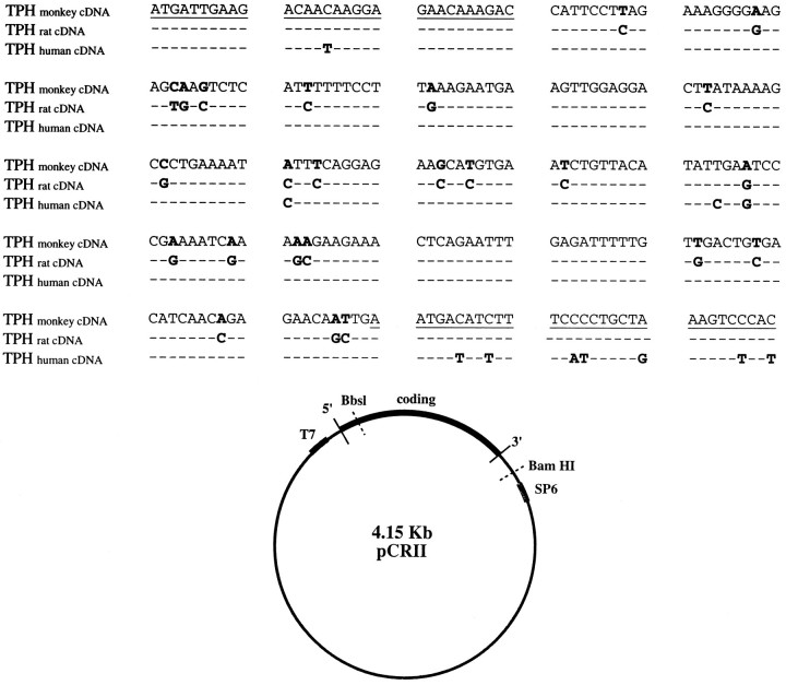
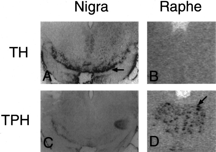
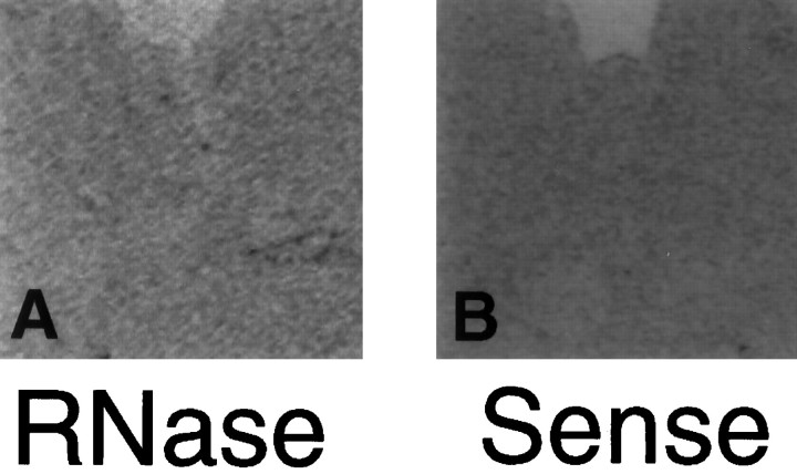
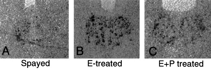
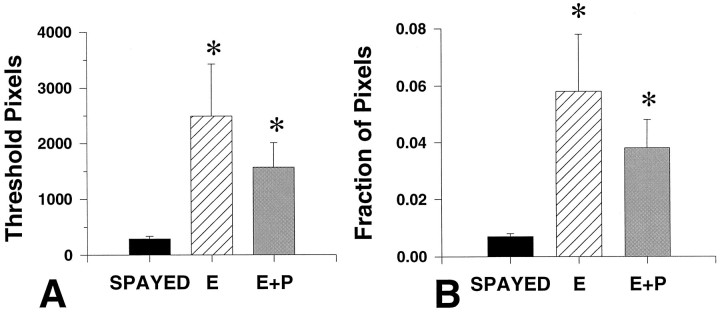
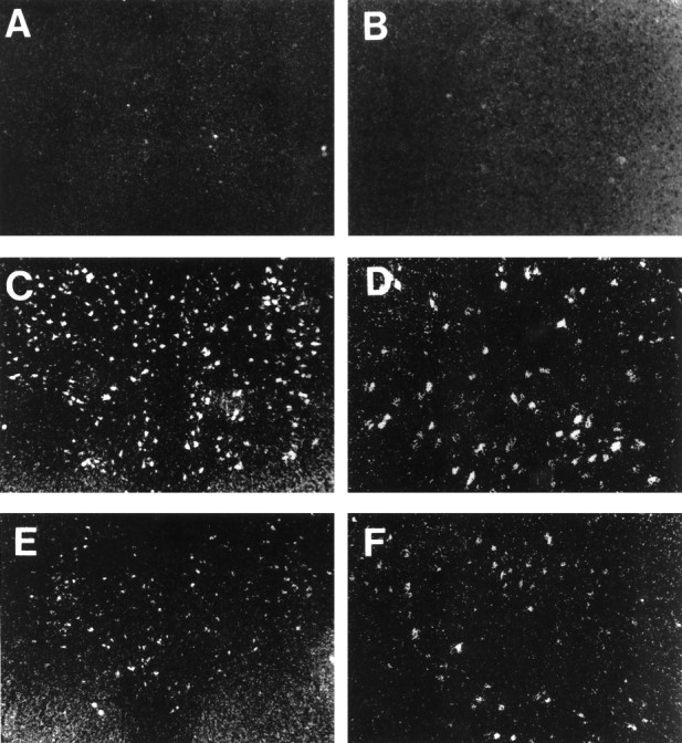
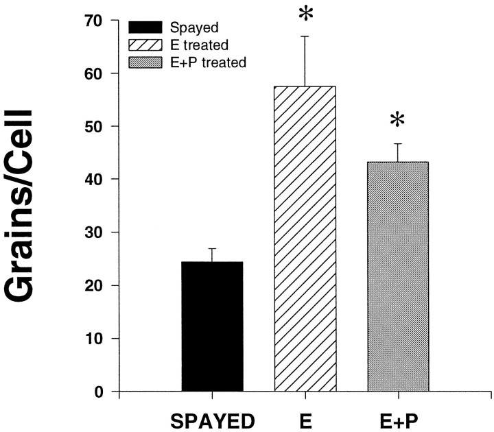
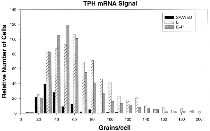
Progesterone (P) stimulates prolactin secretion through an unknown neural mechanism in estrogen (E)-primed female monkeys. Serotonin is a stimulatory neurotransmitter in prolactin regulation, and this laboratory has shown previously that E induces progestin receptors (PR) in serotonin neurons. Therefore, we questioned whether E and/or E+P increased serotonin neural function. The expression of mRNA for tryptophan hydroxylase (TPH) was examined in ovariectomized (spayed) control, E-treated (28 d), and E+P-treated monkeys (14 d E and 14 d E+P) using in situ hybridization and a 249 bp TPH cRNA probe generated with RT-PCR (n = 5 animals/group). Densitometric analysis of film autoradiographs revealed a ninefold increase in TPH mRNA in E-treated macaques compared to spayed animals (p < 0.05). With supplemental P treatment, TPH mRNA signal was increased fivefold over spayed animals (p < 0.05), but was not significantly different compared to E-treated animals. These results were verified by grain counts from photographic emulsion-coated slides. There were significantly higher single-cell levels of TPH mRNA in serotonergic neurons of the dorsal raphe in E- and E+P-treated groups (p < 0.05). These data indicate that E induces TPH gene expression in nonhuman primates and that the addition of P has little additive effect on TPH gene expression. Thus, the action of P on prolactin secretion is probably not mediated at the level of TPH gene transcription. However, because P increases raphe serotonin content in E-primed rodents, the possibility remains that P may have other actions on post-translational processing or enzyme activity.


Figures

Fig. 1.
Fig. 1.
A, Nucleotide sequence of amplified monkey dorsal raphe TPH cDNA compared to rat (Kim et al., 1991) and human TPH (Boularand et al., 1990). The coding sequence is numbered from nucleotide 1. Underlined regions indicate the location of PCR primers. Bold letters indicate differences in the nucleotides between sequences. Nucleotides that are identical to those in monkey TPH appear as dashes.B, Plasmid map of monkey 5′ TPH cDNA showing restriction enzyme recognition and promoter sites. pCR II was the 3.9 kb plasmid used for ligation of the 0.25 kb monkey TPH cDNA. The cDNA clone was oriented for antisense transcription by SP6 RNA polymerase and linerized for transcription via a BbsI digest. Restriction endonuclease BamHI was used to linearize the plasmid for coding sequence. T7 RNA polymerase was then used to make sense cDNA.
Fig. 2.
Fig. 2.
Demonstration of cell specificity for the hybridization of TPH riboprobe in monkey brain. A andC depict sections of substantia nigra. Band D depict sections of raphe. In A andB, TH riboprobe was applied to the substantia nigra (A) and the raphe (B). The TH probe hybridized only to the dopaminergic neurons of the nigra as indicated by the black arrow (A). InC and D, TPH riboprobe was applied to the substantia nigra (C) and the raphe (D). The TPH riboprobe hybridized only to the serotonergic neurons of the raphe as indicated by the black arrow(D). These are computer-generated images (digitized) of scanned autoradiographs, which are not as sharp as photomicrographs obtained directly through a microscope lens.
Fig. 3.
Fig. 3.
Digitized autoradiographs of control sections.A, Antisense TPH riboprobe applied after RNase pretreatment in the raphe produced no hybridization signal.B, No hybridization was observed in the raphe when a sense stand of TPH riboprobe was applied.
Fig. 4.
Fig. 4.
Representative autoradiographs (digitized) from equivalent levels of the dorsal raphe obtained from a spayed, an E-treated, and an E+P-treated monkey. There appeared to be an increase in TPH mRNA signal as reflected by black pixels in the dorsal raphe of both E- and E+P-treated animals.
Fig. 5.
Fig. 5.
A, Average number of above-threshold pixels measured in spayed and steroid-treated animals (n = 5 animals/treatment group) with five levels per animal. B, The fraction of pixels that are the result of specific labeling in the dorsal raphe over total pixel number in spayed, E-treated, and E+P-treated animals.Asterisks, Significantly different from spayed;p < 0.05. There were no significant differences between E- and E+P-treated groups.
Fig. 6.
Fig. 6.
Dark-field photomicrographs of cells of the dorsal raphe that were labeled for TPH mRNA by ISH followed by emulsion development of silver grains. Representative raphes from a spayed (A, B), an E-treated raphe (C, D), and an E+P-treated monkey (E, F) are shown. Magnification:right panels, 12.5×; left panels, 25×.
Fig. 7.
Fig. 7.
Average number of grains per cell in serotonergic dorsal raphe cells from spayed and steroid-treated monkeys (n = 3 animals/spayed group; n = 4 animals/steroid treatment group) with five levels analyzed per animal. There was a significant increase in TPH mRNA with E and E+P.Asterisks, Significantly different from spayed group;p < 0.05. There was no significant difference between E- and E+P-treated groups.
Fig. 8.
Fig. 8.
Frequency distribution of TPH mRNA silver grains in neurons of the dorsal raphe of spayed (n = 3), E-treated (n = 4), and E+P-treated (n = 4) animals. The frequency distribution of the E- and E+P-treated animals is shifted to the right compared with spayed animals, indicating an increase in signal intensity in the steroid-treated groups. E- and E+P-treated groups also had more cells that were positive for TPH mRNA.

References

    1. Anderson IM, Ware CJ, Da Roza Davis JM, Cowen PJ. Decreased 5-HT-mediated prolactin release in major depression. Br J Psychiatry. 1992;160:372–378. - PubMed
    1. Bethea CL. Colocalization of progestin receptors with serotonin in the raphe neurons of macaque. Neuroendocrinology. 1993;57:1–6. - PubMed
    1. Bethea CL. Regulation of progestin receptors in raphe neurons of steroid-treated monkeys. Neuroendocrinology. 1994;60:50–61. - PubMed
    1. Bethea CL, Papkoff H. Purification of monkey prolactin from culture medium: biochemical and immunological characterization. Proc Soc Exp Biol Med. 1986;182:22–33. - PubMed
    1. Bethea CL, Fahrenbach WH, Sprangers SA, Freesh F. Immunocytochemical localization of progestin receptors in monkey hypothalamus: effect of estrogen and progestin. Endocrinology. 1992;130:895–905. - PubMed

Publication types

MeSH terms

Associated data

LinkOut - more resources