. 1996 Nov 1;16(21):7063-76.
doi: 10.1523/JNEUROSCI.16-21-07063.1996.

Localization of rhythmogenic networks responsible for spontaneous bursts induced by strychnine and bicuculline in the rat isolated spinal cord

Affiliations

Localization of rhythmogenic networks responsible for spontaneous bursts induced by strychnine and bicuculline in the rat isolated spinal cord

E Bracci et al. J Neurosci. .

Abstract

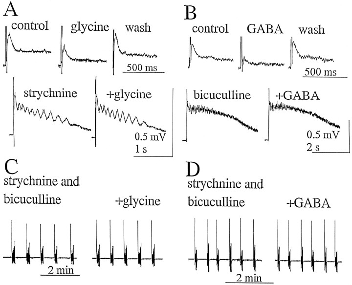
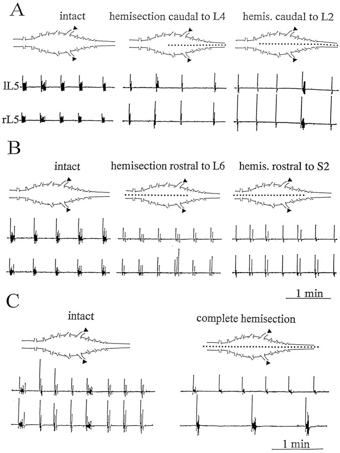
Spontaneous rhythmic bursting induced by coapplication of strychnine (1 microM) and bicuculline (20 microM) was observed with electrophysiological recording from pairs of lumbar ventral roots (usually L5) in an isolated preparation of the neonatal rat spinal cord. Bursting was insensitive to exogenously applied GABA or glycine, confirming that it was attributable to block of glycine and GABAA receptor-mediated inhibition. NMDA accelerated bursting in a dose-dependent manner. Complete coronal spinal transection at L3 or L6 level did not block bursting recorded from L5 or L2 roots, respectively. Gradual cutting of the cord along the midline through a sagittal plane preserved bursting activity in both disconnected sides but led to loss of synchronicity. Once the spinal cord was fully separated into left and right halves, regular bursting persisted on each side with no phase-coupling between the two preparations. Section along a frontal plane to remove dorsal horns and much of the central canal area did not affect burst frequency or left-to-right synchronicity, whereas it reduced burst duration. A quadrant preparation containing mainly a single ventral horn displayed enhanced burst frequency while bursts became very short events. Bath application of 5-hydroxytryptamine (30 microM) or NMDA (5 microM) increased burst frequency and decreased burst duration in all types of preparation except the isolated quadrants, in which brief bursts were accelerated but not shortened by these chemical agents. These results suggest that bursting induced by strychnine and bicuculline apparently relied on distinct mechanisms for burst triggering and intraburst structure. The first required a relatively smaller neuronal network that was confined to a ventral quadrant. Intraburst structure was dependent on a larger circuitry comprising either both ventral horns or one side of the spinal cord including more than two segments.


Figures

Fig. 1.
Fig. 1.
Comparison of intracellular and extracellular recordings of bursting activity induced by strychnine and bicuculline.A, Intracellular traces from L5 motoneuron (resting membrane potential −75 mV) displaying regular rhythmic bursting characterized by rapid depolarization with superimposed trains of action potentials. A single event (indicated by thearrow) is shown on the right with a faster timebase to resolve intraburst oscillatory activity.B, DC-coupled extracellular recording of similar bursting (different preparation from A) from L5 ventral root. Note the similar bursting pattern and structure as observed with the intracellular electrode. Traces on the rightrepresent faster timebase records [with wideband (top) or lowpass (bottom) filter] of the same individual burst indicated by the arrow. C, AC-coupled extracellular recording of the same activity depicted inB. Right traces show the same single event (faster timebase) with wideband (top) or lowpass (bottom) filter. Note that extracellular recording (either DC- or AC-coupled) from ventral root provides a reliable method to observe bursting activity.
Fig. 3.
Fig. 3.
Dose-dependent action of NMDA on bursting activity induced by strychnine and bicuculline. A, Pair of AC-coupled tracings (left and right L5 ventral root; lL5and rL5; 20 Hz lowpass filtering) in strychnine and bicuculline solution (top) and after application of NMDA at the concentrations shown above each trace. Right panels show faster time base records of individual bursts (arrows) to depict intraburst oscillations. Note progressive acceleration of bursting activity by NMDA with concomitant decrease in burst duration. B, Plot of NMDA concentration versus burst duration (top) or cycle period (bottom). Each datapoint was calculated over a period of at least 5 min for the preparation shown inA.
Fig. 4.
Fig. 4.
Effect of cross-sectioning the spinal cord at L3 level on bursting activity. A, AC-coupled sample records of bursting induced by strychnine and bicuculline (see legend to Fig. 3for further details; no 20 Hz filtering) in an isolated preparation (intact) and after removal of the cord region rostral to L3 (see schematic diagram on the left). Right traces are faster records of arrowed events.B, Histograms of cycle period (left) or burst duration (right) calculated from six preparations before (open columns) or after (hatched columns) this type of lesion. In this and all subsequent figures (unless otherwise indicated), data are normalized with respect to control values before the lesion.
Fig. 2.
Fig. 2.
Effects of glycine or GABA on reflex activity and bursting. A, Top, DC records of polysynaptic reflex activity elicited by dorsal root stimulation (0.1 msec duration, 1/8 sec frequency) before (control), during (glycine), or after (wash) application of 0.5 mm glycine. Responses are averaged records of 10 traces. Those in glycine solution are taken 2 min after the start of glycine superfusion; wash indicates 10 min after glycine washout. Bottom, Strychnine (1 μm) enhanced reflex amplitude and induced oscillatory patterns. This effect was not sensitive to glycine (0.5 mm; same preparation as above). B (different preparation from A),Top, DC records of polysynaptic reflex activity elicited as described in A before, during, or after application of 1 mm GABA. Bottom, Bicuculline (20 μm) enhanced reflex activity and prevented the effect of GABA. All responses are averaged records of 10 traces and were recorded in the presence of CGP 52,432 (10 μm) to block GABAB receptors. C, AC-coupled records of spontaneous bursting activity (same preparation as in A) induced by coapplication of strychnine (1 μm) and bicuculline (20 μm); glycine (0.5 mm) has no depressant action, confirming that glycine receptors are effectively blocked. D, Comparable records (same preparation as inB) showing the lack of depressant action of GABA (1 mm) on bursting activity induced by strychnine and bicuculline (in the presence of CGP 52,432), indicating that GABAA receptors are effectively blocked.
Fig. 5.
Fig. 5.
Effects of sagittal hemisectioning of the spinal cord on bursting activity. A, AC-coupled tracings (from left and right L5 ventral roots marked by filled triangles; 20 Hz lowpass filtering) of bursting activity from the isolated preparation (intact; left) and after two types of lesion bisecting the cord from the caudal end up to L4 (middle) or to L2 (right). Schemes of these lesions are shown above corresponding traces. Synchronized bursting (with shorter burst duration) persists after these lesions.B, Similar tracings from a preparation in which the bisectioning was started from the rostral end down to L6 (middle) or S2 (right) as indicated by schemes above traces. Also, in this case bilateral synchronicity of L5 bursting is preserved. C, Example of bursting activity from a preparation before (left) and after (right) complete hemisectioning of the spinal cord. The latter procedure leads to alteration in the frequency of bursting for each root, loss of synchronicity between the left and right ventral roots, and shorter burst duration.
Fig. 6.
Fig. 6.
Histograms of changes in cycle period and burst duration after partial or complete hemisection of the spinal cord.AC represent three lesion protocols, namely, hemisectioning from the caudal end (open columns), from the rostral end (hatched columns), or complete hemisection (cross-hatched columns). Data are averages from four preparations for each protocol. The extent of hemisectioning in A and B is indicated asI, II, III, orIV (see Results for further details): note that when hemisectioning splits a considerable length of the cord (typeII or IV lesions), there is an increase in the SD of the cycle period (reflecting increased variability of burst frequency) even if average values do not differ significantly. Complete hemisectioning (C) largely increases the variability of the cycle periods and significantly (**p < 0.0001) reduces burst duration.
Fig. 7.
Fig. 7.
Effect of ablation of dorsal horns on bursting activity. A, DC-coupled records of bursting activity from L5 ventral roots before (intact;top) and after frontal section (ventral horns only; bottom), which removes dorsal horns and area below midline (see stained section of this preparation inB, either alone or within the schematic contours of the spinal cord). Right traces are shown with a faster time base to depict individual burst (arrows) time course. Note that this procedure shortens burst duration. C, Histograms of normalized cycle periods (left) and burst duration (right) for unlesioned (open columns) or lesioned (hatched columns) preparations (n = 4). Burst duration is significantly decreased (**p < 0.0001).
Fig. 8.
Fig. 8.
Bursting activity in an isolated ventral horn quadrant. A, DC-coupled tracings (from right L5 ventral root) in isolated preparation (intact;top) and after surgical isolation (bottom) of a ventral quadrant. The latter is shown inB histologically either alone or within the idealized contours of the spinal cord. Responses indicated byarrows are shown on the right side ofA on a faster time base. C, Histograms of cycle period (left) or burst duration (right) for unlesioned (open columns) or lesioned (hatched columns) preparations (n = 4). **p < 0.0001.
Fig. 9.
Fig. 9.
Sensitivity of bursting to NMDA in a ventral quadrant preparation. A, DC-coupled tracings of bursting activity from left L5 ventral root in a ventral quadrant preparation (for details, see legend to Fig. 8) in strychnine and bicuculline solution (top) to which NMDA (5 μm) is then added (bottom). Right traces are shown with faster time base. Note increase in burst frequency in the presence of NMDA. B, Histograms of cycle period (in sec;left) or burst duration (right) in strychnine + bicuculline solution (open columns) or after addition of NMDA (hatched columns). Data are means of values observed during 5 min periods. **p < 0.0001.

References

    1. Becker CM, Hoch W, Betz H. Glycine receptor heterogeneity in rat spinal cord during postnatal development. EMBO J. 1988;7:3717–3726. - PMC - PubMed
    1. Ballerini L, Bracci E, Nistri A. Desensitization of AMPA receptors limits the amplitude of EPSPs and the excitability of motoneurons of the rat isolated spinal cord. Eur J Neurosci. 1995;7:1229–1234. - PubMed
    1. Bracci E, Ballerini L, Nistri A. Spontaneous rhythmic bursts induced by pharmacological block of inhibition in lumbar motoneurons of the neonatal rat spinal cord. J Neurophysiol. 1996;75:640–647. - PubMed
    1. Cazalets JR, Sqalli-Houssaini Y, Clarac F. Activation of the central pattern generators for locomotion by serotonin and excitatory amino acids in neonatal rat. J Physiol (Lond) 1992;455:187–204. - PMC - PubMed
    1. Cazalets JR, Borde M, Clarac F. Localization and organization of the central pattern generator for hindlimb locomotion in newborn rat. J Neurosci. 1995;15:4943–4951. - PMC - PubMed

Publication types

MeSH terms

Grants and funding

LinkOut - more resources