. 1996;5(6):301-13.

Analysis of small nuclear RNAs in a precatalytic spliceosome

Affiliations

Analysis of small nuclear RNAs in a precatalytic spliceosome

S L Yean et al. Gene Expr. 1996.

Abstract

U1 small nuclear RNA plays an important role in early stages of intron recognition and spliceosome assembly. The 5' splice site of the premessenger RNA base-pairs with the 5' end of U1; however, that interaction appears to be replaced by U5 and U6 at later stages of the splicing process. It has not been established when this transition occurs nor what factors are required for the transition. The PRP2 gene of Saccharomyces cerevisiae encodes an RNA-dependent ATPase that belongs to the DEAH putative RNA helicase family. A spliceosome can be assembled in the absence of PRP2 but the ATPase activity of PRP2 is required for the onset of the catalytic reactions in the spliceosome. By probing the precatalytic spliceosome formed in temperature-sensitive prp2 mutant extracts with oligonucleotides complementary to snRNAs, we found that the 5' end of U1 was sensitive to RNase H digestion whereas the 5' splice site-interacting region of U6 became resistant. Furthermore, by treating with heparin, a spliceosome depleted of U1 snRNA was isolated that subsequently underwent splicing with additional protein factors and ATP. Thus, these results indicate that PRP2 is not responsible for the transition from U1 to U6 in the spliceosome and that the entire U1 snRNA is not involved in the catalytic phase of pre-mRNA splicing.


Figures
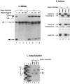
FIG. 1
FIG. 1
Cleaving snRNAs in extracts before or after spliceosome assembly by oligonucleotide-directed RNase H digestion. Three oligonucleotides, 1s19, 2b19, and 6c27, complementary to U1, U2, and U6 snRNA, respectively, were used to target specific snRNA cleavage by the endogenous RNase H. For cleaving before spliceosome formation, oligonucleotides were incubated with the prp2 Ts extracts prior to the addition of pre-mRNA substrate; for cutting afterwards, spliceosomes containing pre-mRNA were allowed to be assembled in the Ts extracts followed by the addition of oligonucleotides. Purified wild-type PRP2 protein was added last to complete the splicing reactions. In all figures, lanes of gels are numbered at the bottom of the autoradiograms. (A) Splicing activity assays of reactions containing 32P-labeled pre-mRNA substrates on denaturing polyacrylamide gels. Lanes 1–4: oligonucleotides were added prior to the assembly of the spliceosome; lanes 5–8: oligonucleotides were added after spliceosome assembly. Water, instead of oligonucleotide, was used in samples in lanes 1 and 5 as control. (B) Northern analysis of the cleavage events. RNA was recovered from reaction mixtures containing unlabeled pre-mRNA, separated on three gels (polyacrylamide gels for U1 and U6, agarose gel for U2), and probed with U1 (lanes 1–4), U2 (lanes 5–8), and U6 (lanes 9–12). No oligonucleotides were used in samples in odd-number lanes; samples in even-number lanes were treated with 1s19 (lanes 2 and 4), 2b19 (lanes 6 and 8), or 6c27 (lanes 10 and 12). (C) Mapping the cleavage sites on U1 snRNA by primer extension. RNA was recovered from samples treated with water (lanes 6 and 8) or with oligonucleotide 1s19 (lanes 5 and 7) and was reverse-transcribed with a 32P-labeled oligonucleotide complementary to the 74th to 92nd bases of U1 (Table 1). Dideoxy sequencing reactions on U1 DNA were used as size markers (lanes 1–4).
FIG. 2
FIG. 2
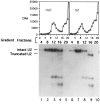
Gradient and Northern analyses of U2 snRNA after oligonucleotide-directed RNase H digestion. Oligonucleotide 2b19 was added to initiate RNase H digestion after the assembly of the spliceosome in prp2 Ts extracts. The reaction mixtures were treated with heparin at 0.5 μg/μl, then sedimented through glycerol gradients. The graphs show the gradient profiles (fractions with smaller numbers were closer to the bottom of the gradient) of the control reaction containing 32P-labeled pre-mRNA without oligonucleotide (H2O) and the 2b19-treated, radioactive sample (U2). Total RNA was isolated from “cold” gradient fractions 4, 8, 12, 16, and 20 (water control, lanes 1–5; 2b19-treated, lanes 6–10), separated on an agarose gel, and probed with U2. Fraction numbers are given on the top of the autoradiogram and lanes are numbered at the bottom.
FIG. 3
FIG. 3
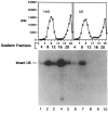
Gradient and Northern analyses of U6 snRNA after oligonucleotide-directed RNase H digestion. Oligonucleotide 6c27 was added to initiate RNase H digestion after the assembly of the spliceosome in prp2 Ts extracts. The “hot” gradient profiles and the Northern hybridization using “cold” fractions probed with U6 were displayed as in Fig. 2.
FIG. 4
FIG. 4
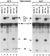
Gradient and Northern analyses of U1 snRNA after oligonucleotide-directed RNase H digestion. Oligonucleotide 1s27 or 1s19 was added to initiate RNase H digestion after the assembly of the spliceosome in prp2 Ts extracts. The reaction mixtures were treated with heparin at 0.5 μg/μl (water control and 1s27-treated samples) or 1.25 μg/μl (H19-treated sample) prior to centrifugation. The “hot” gradient profiles are displayed above the Northern autoradiograms: the top was probed with actin DNA (exposed to an X-ray film for 49 h with two LP screens), the middle probed with U1 DNA (78.5 h with two LP screens), and the bottom was probed with U1 plus actin sequences (25.5 h with one LP screen). Lanes 1–5: water control; lanes 6–10: 1s27-treated sample; lanes 11–15: 1s19-treated sample containing high concentration of heparin; lane 16: purified PRP2 protein; lane 17: purified HP factor. See Fig. 2 for additional description.
FIG. 5
FIG. 5
Splicing activity assays of the gradient-purified spliceosomes treated with U2 (2b19) or U6 (6c27) oligonucleotide as shown in Figs. 2 and 3. The “hot” spliceosome-containing fractions were incubated with Buffer D (lanes 1, 4, 7, 10), wild-type extracts (lanes 2, 5, 8, 11), or two extrinsic splicing factors, PRP2 and HP (lanes 3, 6, 9, 12) under splicing conditions. After the incubation RNA was analyzed on sequencing gels followed by autoradiography. Lanes 1–3 and 7–9: water control spliceosomes; lanes 4–6: 2b19-digested spliceosomes; lanes 10–12: 6c27-digested spliceosomes.
FIG. 6
FIG. 6
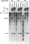
Splicing activity assays of the gradient-purified spliceosomes treated with U1 oligonucleotides as shown in Fig. 4. The “hot” spliceosome-containing fractions were assayed as described in Fig. 5. Lanes 1–3: water control spliceosomes; lanes 4–6: H27-digested spliceosomes; lanes 7–9: 1s19-digested spliceosomes treated with a higher concentration of heparin. Note that the longer EcoRI transcript was used in this experiment.

Similar articles

Cited by

References

    1. Abelson J. RNA processing and the intervening sequence problem. Annu Rev. Biochem. 48:1035–1069; 1979. - PubMed
    1. Brody E.; Abelson J. The spliceosome: Yeast pre-messenger RNA associates with a 40S complex in a splicing-dependent reaction. Science 228:963–967; 1985. - PubMed
    1. Cech T. R. The generality of self-splicing RNA: Relationship to nuclear mRNA splicing. Cell 44:207–210; 1986. - PubMed
    1. Cheng S. C.; Abelson J. Spliceosome assembly in yeast. Genes Dev. 1:1014–1027; 1987. - PubMed
    1. Company M.; Arenas J.; Abelson J. Requirement of the RNA helicase-like protein PRP22 for release of messenger RNA from spliceosomes. Nature 349:487–493; 1991. - PubMed

Publication types

LinkOut - more resources