. 1996;5(6):315-30.

Tissue specificity of L-pyruvate kinase transgenes results from the combinatorial effect of proximal promoter and distal activator regions

Affiliations

Tissue specificity of L-pyruvate kinase transgenes results from the combinatorial effect of proximal promoter and distal activator regions

L Miquerol et al. Gene Expr. 1996.

Abstract

The L-type pyruvate kinase (L-PK) gene is regulated by diet and hormones and expressed at high levels in the hepatocytes, enterocytes, and proximal tubular cells of the kidney and at low levels in the endocrine pancreatic cells. Two regulatory regions have been shown to be important in transgenic mice to confer on a reporter gene a similar tissue-specific and diet-responsive expression: a proximal promoter fragment, with binding sites for the tissue-specific hepatocyte nuclear factors 1 and 4, and presence of the glucose-response element (GIRE) and a distal activator corresponding to a liver-specific hypersensitive site at -3000 bp with respect to the cap site. Although the proximal promoter is able to confer by itself tissue-specific expression on a reporter gene, its activity in vivo is strongly stimulated by the distal activator. To determine the possible role of the distal region on diet responsiveness and tissue specificity of the L-PK gene expression, we have created lines of transgenic mice in which the gene for SV40 T antigen (Tag) was directed by composite regulatory sequences consisting of the L-PK promoter and different enhancers: either the SV40 early enhancer (SV) or the H enhancer of the aldolase A gene (H). The induction of the composite H-PK/Tag and SV-PK/Tag transgenes by a carbohydrate-rich diet in the liver was similar to that of the endogenous L-PK gene. This suggests that in fasted mice the L-PK promoter, and especially the GIRE, is able to silence the activating influence of a strong viral enhancer such as the SV40 enhancer. The H-PK/Tag mice expressed the transgene similarly to the endogenous gene, except in the pancreas, where expression was practically undetectable. Consistently, whereas L-PK/Tag mice develop insulinomas, H-PK/Tag mice develop only hepatomas. In contrast, the transgene expression was partly aberrant in SV-PK/Tag mice. In addition to a normal activation of the transgene in the liver, a strong expression was also detected in the kidney medulla, whereas the transgene was practically silent in enterocytes. Finally, the effect of the distal region (-2070 to -3200) on an ubiquitous promoter was tested by ligating the distal L-PK gene fragment in front of a thymidine kinase/CAT transgene. Such a transgene was constantly expressed in the pancreas and, strikingly, in the brain. It appears, therefore, that the L-PK distal activator exhibits, by itself, a certain neuropancreatic specificity required in combination with the proximal promoter for L-PK gene expression in pancreas endocrine cells.


Figures

FIG. 1
FIG. 1
Structure of the hybrid pyruvate kinase transgenes. The L-PK/Tag transgene was composed of 2.7 kbp of the SV40 t and T antigens sequences (hatched rectangles) directed by 3.2 kbp of 5′ flanking regions of the rat L-PK gene (7,10). The black rectangles represent the erythroid (L′) and liver (L) specific exons of the L-PK gene. The H-PK/Tag and SV-PK/Tag were created by excision of 2.2 kbp of L-PK regulatory sequences between −3200 and −1000 nt of the L-PK/Tag clone. This region was replaced by enhancer H of aldolase A (H-PK/Tag) or SV40 enhancer (SV-PK/Tag). The designation of each mouse line and corresponding copy number are given on the right part of the figure.
FIG. 2
FIG. 2
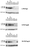
Northern blot analysis of transgene expression in various tissues. Northern blot analyses were performed with 10 μg of total RNAs from the liver, kidney, intestine, and spleen of transgenic mice fed a high-carbohydrate diet. The membranes were successively hybridized with Tag specific probe and with rRNA (18S) probe (R45) used as a standard (12). Mouse lines are indicated above the panels: (A) L-PK/Tag and H-PK/Tag lines and (B) L-PK/Tag and SV-PK/Tag lines.
FIG. 3
FIG. 3
RT-PCR analysis of L and L′-PK/Tag transcripts in the different types of transgenic mice. Total RNAs from 500 ng of various tissues were used for reverse transcriptase PCR amplifications. The L-PK-Tag amplified fragment is 490 bp long and the L′-PK-Tag amplified fragment is 550 bp long. The mouse β-actin specific amplified fragment is 241 bp long and served as standard. Southern blotting of poly-acrylamide gel and hybridization were described in the Materials and Methods section.
FIG. 4
FIG. 4
Northern blot analysis of transgene dietary regulation. Northern blot analyses were performed with 10 μg of total RNAs from liver, kidney, intestine, and spleen of transgenic mice fasted for 24 h (F) or refed a high-carbohydrate diet (75% of glucides) (R). We used 2.7 kb of SV 40 T-antigen sequences to reveal the transgene expression (t, T antigens) and the 18S R45 probe as a standard.
FIG. 5
FIG. 5
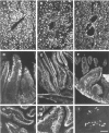
Nuclear T antigen immunolocalization in livers and intestines from transgenic mice. Frozen sections from livers (A-C) and small intestines (D-I) of L-PK/Tag1 (A, D, G), H-PK/Tag38 (B, E, H), and SV-PK/Tag12 (C, F, I) transgenic mice were processed for the immunodetection of Tag using a specific polyclonal antibody as described in the Materials and Methods section. In the three lines of transgenic mice, 100% of liver cells exhibited a nuclear large T antigen positivity (A-C). Note the presence of numerous mitotic cells in the liver of H-PK/Tag transgenic mice (C). In the small intestine the pattern of labeling differed in the three lines of transgenic mice. In both L-PK/Tag1 and H-PK/Tag38 transgenic mice, an intense nuclear labeling was observed in enterocytes lining the crypt/villus axis (D, E, G, H). In contrast, the nuclear labeling was almost not detectable in small intestine sections of SV-PK/Tag12 transgenic mice (F, I)). Bars = 50 μm.
FIG. 6
FIG. 6
Nuclear T antigen immunolocalization in kidneys and pancreas from transgenic mice. Frozen sections from kidney cortex (A-C), kidney medulla (D-F), and pancreas (G-I) of L-PK/Tag1 (A, D, G), H-PK/Tag38 (B, E, H), and SV-PK/Tag12 (C, F, I) transgenic mice were processed for the immunodetection of Tag using a specific polyclonal antibody as described in the Materials and Methods section. In kidney cortex the nuclear labeling was restricted to the proximal tubule cells in the three lines of trangenic mice (A, B, C). Glomeruli (arrowheads) and distal tubules (arrows) were not stained. No labeling was detected in the kidney medulla of L-PK/Tag1 (D) and H-PK/Tag38 (E) transgenic mice. In contrast, the nuclei from most tubules sections were labeled in kidney medulla of SV-PK/Tag12 mice (F). The pattern of labeling also varied in the pancreas from the various lines of transgenic mice. In L-PK/Tag1 mice (G), all nuclei from exocrine and endocrine (arrowhead) pancreas were labeled. Note that the nuclear labeling was more intense in the endocrine than in the exocrine pancreatic cells. Endocrine pancreatic cells from both H-PK/Tag38 (H) and SV-PK/Tag12 (I) transgenic mice also presented a positive nuclear labeling (arrowheads), whereas exocrine pancreatic cells were almost not labeled (H, I). Bars = 50 μm.
FIG. 7
FIG. 7
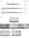
Tissue-specific expression of HSS2-TKCAT constructs in transgenic mice. (A) Structure of the HSS2-TK/ CAT hybrid gene. HSS2-TK/CAT and HSS2b-TK/CAT were obtained by the insertion of a 1111-bp-long fragment encompassing the HSS2 site in both orientation (hatched boxes), upstream of the thymidine kinase promoter from HSV spanning nt −105 to +51 and the CAT structural gene-SV40 poly(A) cassette (dot boxes). The designation of each mouse line and the corresponding copy number are given on the right part of the figure. (B) Tissue-specific expression of HSS2-TK/CAT constructs in transgenic mice. Samples from homogenates of the indicated organs assayed for CAT activity; 300 μg of proteins was used. The line numbers are indicated below each autoradiograph. Li, liver; P, pancreas; K, kidney; I, small intestine; S, spleen; B, brain; Lu, lung; C, cerebellum.

Similar articles

Cited by

References

    1. Baumhueter S.; Mendel D. B.; Conley P. B.; Kuo C. J.; Turk C.; Graves M. K.; Edwards C. A.; Courtois G.; Crabtree G. R. HNF-1 shares three sequence motifs with the POU domain proteins and is identical to LFB1 and APF. Genes Dev. 4:372–379; 1990. - PubMed
    1. Bergot M.-O.; Diaz-Guerra M.-J. M.; Puzenat N.; Raymondjean M.; Kahn A. Cis-regulation of the L-type pyruvate kinase gene promoter by glucose, insulin and cyclic AMP. Nucleic Acids Res. 20:1871–1878; 1992. - PMC - PubMed
    1. Boquet D.; Vaulont S.; Tremp G. L.; Ripoche M.-A.; Daegelen D.; Jami J.; Kahn A.; Raymondjean M. DNaseI hypersensitivity analysis of the L-type pyruvate kinase gene in rats and transgenic mice. Eur. J. Biochem. 207:13–21; 1992. - PubMed
    1. Botteri F. M.; van der Putten H.; Wong D. F.; Sauvage C. A.; Evans R. M. Unexpected thymic hyperplasia in transgenic mice harboring a neuronal promoter fused with simian virus 40 large T antigen. Mol. Cell. Biol. 7:3178–85; 1987. - PMC - PubMed
    1. Briegel K.; Lim K. C.; Plank C.; Beug H.; Engel J. D.; Zenke M. Ectopic expression of a conditional GATA-2/estrogen receptor chimera arrests erythroid differentiation in a hormone-dependent manner. Genes Dev. 7:1097–1109; 1993. - PubMed

Publication types

MeSH terms

LinkOut - more resources