Case Reports
. 1996 Apr;5(4):517-24.
doi: 10.1093/hmg/5.4.517.

Breakage in the SNRPN locus in a balanced 46,XY,t(15;19) Prader-Willi syndrome patient

Affiliations
Case Reports

Breakage in the SNRPN locus in a balanced 46,XY,t(15;19) Prader-Willi syndrome patient

Y Sun et al. Hum Mol Genet. 1996 Apr.

Abstract

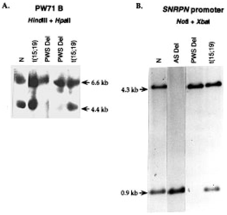
A patient with Prader-Willi syndrome (PWS) was found to carry a de novo balanced reciprocal translocation, t(15;19)(q12;q13.41), which disrupted the small nuclear ribonucleoprotein N (SNRPN) locus. The translocation chromosome 15 was found to be paternal in origin. Uniparental disomy and abnormal DNA methylation were ruled out. The translocation breakpoint was found to have occurred between exon 0 (second exon) and 1 (third exon) of the SNRPN locus outside of the SmN open reading frame (ORF), which is intact. The transcriptional activities of ZNF127, IPW, PAR-1, and PAR-5 were detected with RT-PCR from fibroblasts of the patient, suggesting that these genes may not play a significant role in the PWS phenotype in this patient. Transcription from the first two exons and last seven exons of the SNRPN gene was also detected with RT-PCR; however, the complete mRNA (10 exons) was not detected. Thus, the PWS phenotype in the patient is likely to be the result of disruption of the SNRPN locus.


Figures

Figure 1
Figure 1
Front facial view of the proband (3 years and 6 months old). Note narrow bifrontal diameter, almond-shaped eyes, and down-turned mouth.
Figure 2
Figure 2
Chromosome GTG high resolution banding (top) showing 46,XY,t(15;19)(q12;q13.41). Only the chromosomes 15 and 19 are shown. The ideogram (bottom) shows the translocation breakpoint indicated by arrows.
Figure 3
Figure 3
Parental origin of the translocation chromosome. The normal chromosome 15 in the patient is inherited from his mother (C). The der(15) chromosome is inherited from the father (B). One of the chromosomes 15 of the father of the patient showed medium intensity centromere staining with a pale satellite. The other chromosome 15 in the father showed light centromere staining with a pale satellite. One of the mother’s chromosome 15 showed a pale centromere and a bright satellite. The other chromosome 15 in the mother showed medium to pale centromere staining and a negative satellite. The patient’s normal chromosome 15 showed a pale centromere and bright satellite, indicating it is maternal in origin. The der(15) showed a light centromere and pale satellite, indicating it is paternal in origin.
Figure 4
Figure 4
DNA methylation analysis. N, normal individual; t(15;19), the translocation patient described in this paper; PWS del, PWS deletion patient; AS del, Angelman deletion patient. (A) Leucocyte DNA was digested with HindIII and HpaII and hybridized with the PW71 B probe. The lower band (4.7 kb) is not detected in PWS deletion patients, but is present in normal individuals and the t(15;19) translocation patient. (B) The probe used was a cloned fragment from the SNRPN exon −1 region (16). DNA was digested with NotI and XbaI. The lower band is undetectable in PWS deletion patients, and for AS patients, the upper band is undetectable. Both bands are detectable in normal individuals and the t(15;19) translocation patient.
Figure 5
Figure 5
FISH analysis. (A) Cosmid probe SNRPN; (B) D15S11; (C) GABRB3; (D) D15S10; (E) SNRPN subfragment F2 (Fig. 6A); (F) SNRPN subfragment F3 (Fig. 6A). In A–D, the labeled probes were purchased from Oncor, and a control locus PML cosmid probe was also included for identification of chromosome 15. It produces signals on 15q22. Arrows indicate der(15) (large arrow), der(19) (small arrow), or normal chromosome 15 (arrow head).
Figure 6
Figure 6
Identification of the translocation breakpoint. (A) Restriction map showing area around the breakpoint. Restriction analysis of SNRPN cosmids A and B showed the contig contained the entire SNRPN gene. Each cosmid produced four subfragments larger than 5 kb after digestion with EcoRI (F1–F6). The normal restriction map around the breakpoint is shown at the bottom (5,16). Fragment F2 (and F1, F5) hybridizes to the der(15), while F3 (and F4, F6) hybridizes to the der(19). The arrow and oblong indicate the breakpoint region, and the ten exons are indicated by numbers −1 through 8. (B, BamHI; N, NotI; E, EcoRI; H, HindIII restriction sites). (B, C) Southern analysis for identification of the t(15;19) translocation breakpoint. DNA was digested with BamHI and hybridized with a cDNA probe (B) containing SNRPN exons 1–8 and a cDNA probe (C) containing exons −1, 0, and 1 (16). Lane 1, father; 2 and 7, t(15;19) proband; 3, mother; 4 and 6, PWS deletion patients; 5, PWS UPD patient; arrow, a novel fragments of 20 kb.
Figure 7
Figure 7
RNA expression analysis with RT-PCR. (A) RT-PCR analysis of ZNF127, IPW, PAR-5, and PAR-1. The amplification was present in RT+ reactions (lane 4), but not in RT reactions (lane 3). Lane 1, normal human brain mRNA for positive control; Lane 2, DNA derived from the translocation patient serving as positive control. It was omitted for IPW since RT-PCR spanned an intron. Lane 3, RNA derived from fibroblasts of the patient was incubated without reverse-transcriptase as control (RT); Lane 4, RNA derived from the patient was incubated with reverse-transcriptase (RT+). (B) RT-PCR analysis for SNRPN exons proximal and distal to the translocation breakpoint. The amplification was present in RT+ reactions (lane 3), but not in RT reactions (lane 2). Lane 1, RT-PCR product from mRNA derived from a normal human brain; Lane 2, RT-PCR product from RNA derived from the patient’s fibroblast culture without reverse-transcriptase (RT); Lane 3, reaction product from RNA derived from the patient’s fibroblast culture with reverse-transcriptase (RT+).
Figure 8
Figure 8
Model for expression of SNRPN exons proximal and distal to the translocation. The maternal SNRPN gene is inactive because of imprinting, and therefore no mRNA from exon −1 to 8 is produced (a). The translocation breakpoint maps between exon 0 and 1, and exons 1 to 8 from chromosome 15 distal to the breakpoint were translocated to chromosome 19 near q13.41 (b, right) forming the der(19). The region of chromosome 19 distal to the chromosome 19 breakpoint was translocated to chromosome 15 forming the der(15) (b, at left). The translocation may also have disrupted a gene on chromosome 19, thus forming two fusion genes (c). The promoter for the SNRPN gene remains active and drives transcription of exon −1 to 0, either extending into the putative chromosome 19 gene or terminating randomly (not shown). This product was detected by RT-PCR with exon −1 and 0 primers (Fig. 8B). The promoter for the putative chromosome 19 gene is also active in fibroblasts and transcription generates a fusion mRNA with SNRPN exons 1–8 that can be detected by RT-PCR with primers from exon 2 and 8 (Fig. 8B). An, polyadenylation signal.

References

    1. Butler MG. Prader-Willi syndrome: current understanding of cause and diagnosis. Am J Med Genet. 1990;35:319–332. - PMC - PubMed
    1. Ledbetter DH, Riccardi VM, Airhart SD, Strobel RJ, Keenan BS, Crawford JD. Deletions of chromosome 15 as a cause of the Prader-Willi syndrome. N Engl J Med. 1981;304:325–329. - PubMed
    1. Butler MG, Palmer CG. Parental origin of chromosome 15 deletion in Prader-Willi syndrome. Lancet. 1983;i:1285–1286. - PMC - PubMed
    1. Mascari MJ, Gottlieb W, Rogan PK, Butler MG, Waller DA, Armour JA, Jeffreys AJ, Ladda RL, Nicholls RD. The frequency of uniparental disomy in Prader-Willi syndrome. Implications for molecular diagnosis. N Engl J Med. 1992;326:1599–1607. - PMC - PubMed
    1. Buiting K, Saitoh S, Gross S, Dittrich B, Schwartz S, Nicholls RD, Horsthemke B. Inherited microdeletions in the Angelman and Prader-Willi syndromes define an imprinting centre on human chromosome 15. Nature Genet. 1995;9:395–400. - PubMed

Publication types

MeSH terms

LinkOut - more resources