. 1996 Sep 26;383(6598):350-4.
doi: 10.1038/383350a0.

Neutralizing antibody to human rhinovirus 14 penetrates the receptor-binding canyon

Affiliations

Neutralizing antibody to human rhinovirus 14 penetrates the receptor-binding canyon

T J Smith et al. Nature. .

Abstract

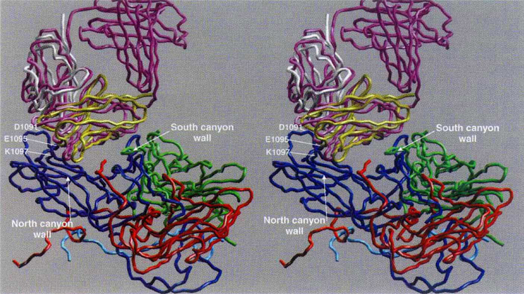
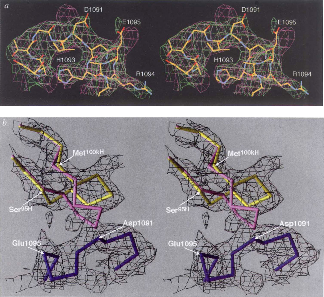
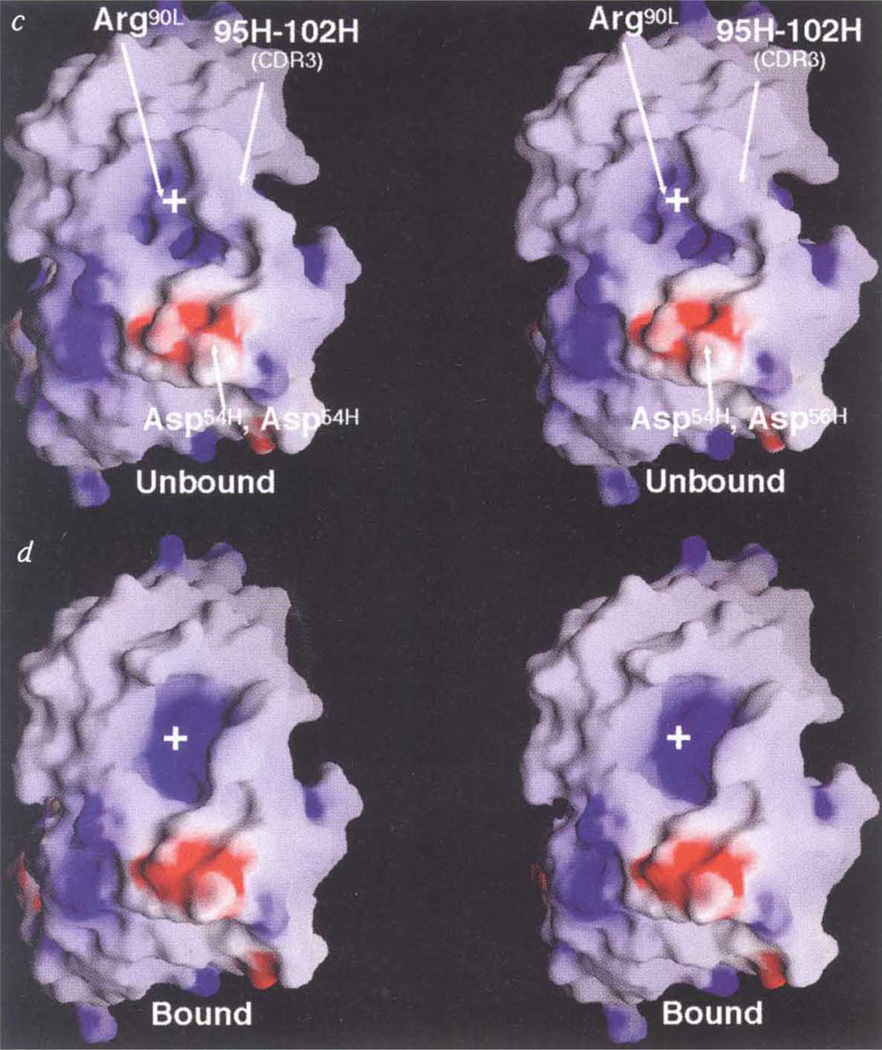
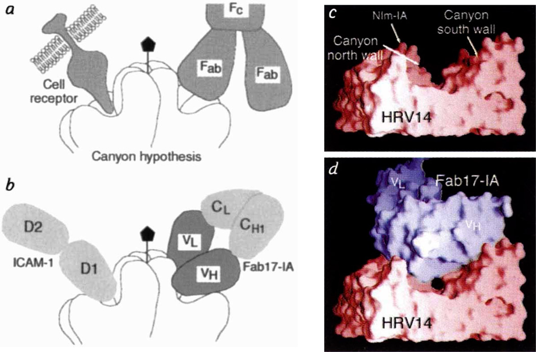
The three-dimensional structure of intact human rhinovirus 14 (HRV-14) complexed with Fab fragments (Fab17-IA) from a strongly neutralizing antibody that binds bivalently to the virion has been determined to 4.0 angstrom resolution by a combination of X-ray crystallography and cryo-electron microscopy. In contradiction to the most commonly held model of antibody-mediated neutralization, Fab17-IA does not induce a conformational change in the HRV-14 capsid. Instead, the paratope of the antibody undergoes a large conformational change to accommodate the epitope. Unlike any previously described antibody-antigen structure, the conserved framework region of the antibody makes extensive contact with the viral surface. Fab17-IA penetrates deep within the canyon in which the cellular receptor for HRV-14 binds. Hence, it is unlikely that viral quaternary structure evolves merely to evade immune recognition. Instead, the shape and position of the receptor-binding region on a virus probably dictates receptor binding and subsequent uncoating events and has little or no influence on concealing the virus from the immune system.


Figures

Figure 1
Figure 1
Cα backbone of the initial and final models. VP1 is blue, VP2 green, VP3 red, initial Fab purple, final VL domain white, and final VH domain yellow. The RNA interior of the virus is towards the bottom of the figure, and some key contact residues around the HRV14 Nlm-IA site are labelled.
Figure 2
Figure 2
Conformational changes at the paratope/epitope interface. a, Comparison between Nlm-IA loop region in the unliganded HRV-14 (mauve) and the Fab/virus complex (green) electron density maps. The atomic coordinates of the native HRV-14 structure were used to calculate the unliganded HRV-14 electron density map, whereas the electron density map of the virus–Fab complex was calculated using phases from 20-fold real-space averaging. The structure of the liganded structure is coloured according to atom type (carbon, yellow; oxygen, red; nitrogen, blue). b, Initial (unliganded Fab structure) and final structures of the heavy-chain CDR3 are purple and yellow, respectively. The Nlm-IA site is shown in dark blue. The black cage is the 4Å electron density map calculated using the experimentally determined phases from 20-fold real-space averaging and phase extension. For this diagram, the VH domains of the initial and final models were aligned to each other. c, d, Stereo diagram of electrostatic potential and structural changes in the hypervariable region before (c) and after (d) binding to the Nlm-IA site. The view is towards the hypervariable region, with the VL domain towards the top of the diagram and the VH domain towards the bottom. Surfaces of positive and negative potential are depicted blue and red, respectively. The largest conformational changes occur in the heavy-chain CDR3 loop and in the side chain of Arg91L. White crosses provide points of reference in the stereo views.
Figure 2
Figure 2
Conformational changes at the paratope/epitope interface. a, Comparison between Nlm-IA loop region in the unliganded HRV-14 (mauve) and the Fab/virus complex (green) electron density maps. The atomic coordinates of the native HRV-14 structure were used to calculate the unliganded HRV-14 electron density map, whereas the electron density map of the virus–Fab complex was calculated using phases from 20-fold real-space averaging. The structure of the liganded structure is coloured according to atom type (carbon, yellow; oxygen, red; nitrogen, blue). b, Initial (unliganded Fab structure) and final structures of the heavy-chain CDR3 are purple and yellow, respectively. The Nlm-IA site is shown in dark blue. The black cage is the 4Å electron density map calculated using the experimentally determined phases from 20-fold real-space averaging and phase extension. For this diagram, the VH domains of the initial and final models were aligned to each other. c, d, Stereo diagram of electrostatic potential and structural changes in the hypervariable region before (c) and after (d) binding to the Nlm-IA site. The view is towards the hypervariable region, with the VL domain towards the top of the diagram and the VH domain towards the bottom. Surfaces of positive and negative potential are depicted blue and red, respectively. The largest conformational changes occur in the heavy-chain CDR3 loop and in the side chain of Arg91L. White crosses provide points of reference in the stereo views.
Figure 3
Figure 3
Fab/virus canyon interactions. a, The HRV/ICAM and HRV/Fab17-IA complexes, showing the HRV-14 canyon that is accessible to cellular receptor but inaccessible to antibody (adapted from ref. 30). b, Current view of virus/receptor and virus/antibody interactions showing that the Fab contacts a large portion of the canyon but not residues at the very bottom (the ‘floor’). Molecular surfaces of HRV-14 (c) and the Fab17-IA/HRV-14 complex(d). The view is parallel to the canyon floor with an icosahedral 5-fold axis towards the left the nearest 2-fold axis towards the right, and the RNA interior towards the bottom of the diagram.HRV-14, the variable domains of the bound Fab, and the molecular interfaces are shown in red, blue, grey, respectively. Only a small portion of the canyon floor is not contacted by the bound Fab.

References

    1. Mosser AG, Leippe DM, Rueckert RR. Molecular Aspects of Picomavirus Infection and Detection. In: Semler BL, Ehrenfeld E, editors. Am. Soc. Microbiol. Washington, DC: 1989. pp. 155–167.
    1. Smith TJ, Olson NH, Cheng RH, Chase ES, Baker TS. Proc. Natl Acad. Sci. USA. 1993;90:7015–7018. - PMC - PubMed
    1. Rossmann MG, et al. Nature. 1985;317:145–153. - PubMed
    1. Olson NH, et al. Proc. Natl Acad. Sci. USA. 1993;90:507–511. - PMC - PubMed
    1. Smith TJ, et al. J. Virol. 1993;67:1148–1158. - PMC - PubMed

Publication types

MeSH terms