

Selective death of hippocampal CA3 pyramidal cells with mossy fiber afferents after CRH-induced status epilepticus in infant rats
- PMID: 8852375
- PMCID: PMC3413264
- DOI: 10.1016/0165-3806(95)00183-2
Selective death of hippocampal CA3 pyramidal cells with mossy fiber afferents after CRH-induced status epilepticus in infant rats
Abstract
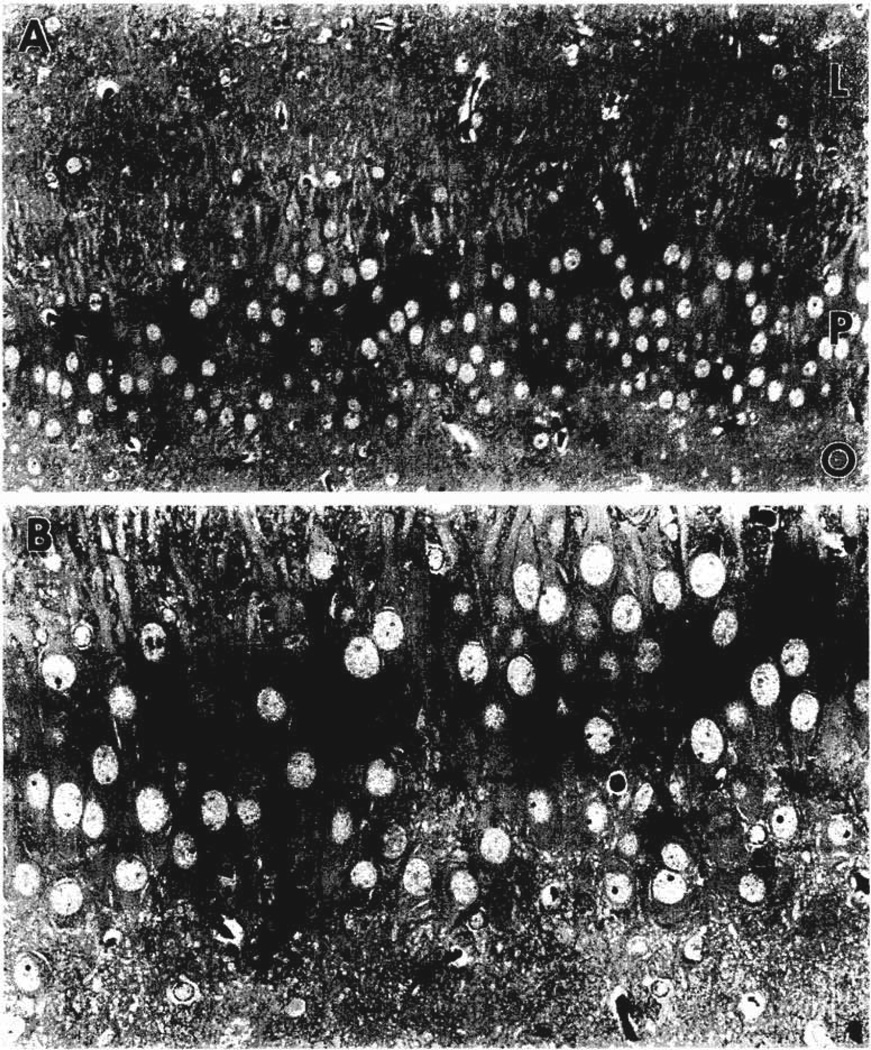
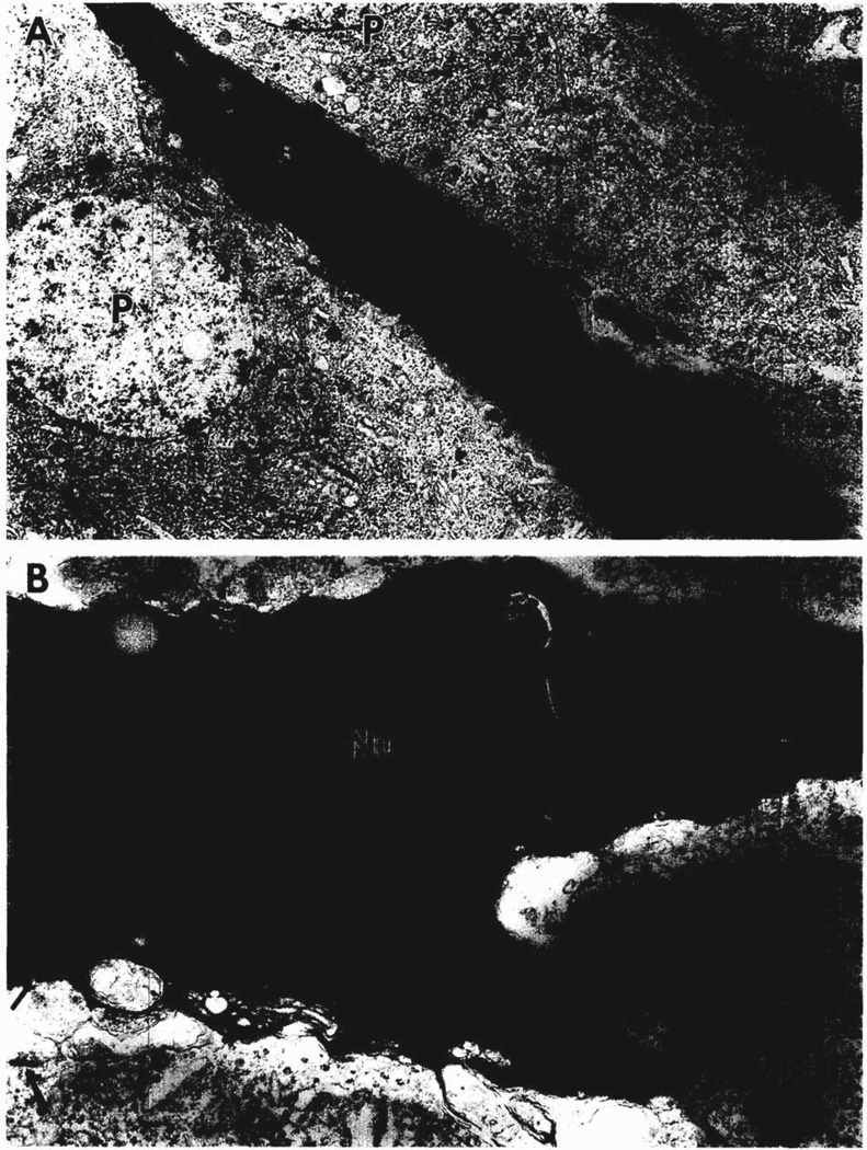
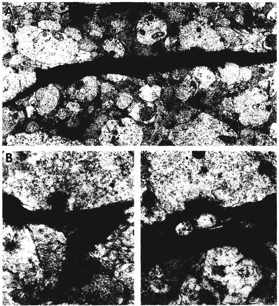
Previous studies of CRH-induced status epilepticus in infant rats demonstrated neuronal loss in several limbic structures, including the CA3 region of the hippocampus. The goal of the present study was to identify the neurons affected by CRH-induced seizures and determine whether they formed synapses with afferent axon terminals. Clusters of neurons in the CA3 region of the hippocampus were osmiophilic when viewed in thick sections. Semi-thin 2-microns sections of the pyramidal cell layer contained dark, shrunken neurons with apical and basal dendrites among normal appearing pyramidal cells. Electron microscopy revealed degenerating pyramidal cells with intact cell membranes and electron dense nuclei and cytoplasm. The shrunken dendrites of these cells had spines and were postsynaptic to large immature-appearing mossy fibers. Thus, CA3 pyramidal neurons that are linked via mossy fibers to the tri-synaptic excitatory hippocampal circuit die subsequent to CRH-induced status epilepticus. The shrunken appearance and selective loss of these neurons are incompatible with necrosis as the mechanism of degeneration.
Figures
Similar articles
-
Postnatal development of CA3 pyramidal neurons and their afferents in the Ammon's horn of rhesus monkeys.Hippocampus. 1995;5(3):217-31. doi: 10.1002/hipo.450050308. Hippocampus. 1995. PMID: 7550617
-
The pro-convulsant actions of corticotropin-releasing hormone in the hippocampus of infant rats.Neuroscience. 1998 May;84(1):71-9. doi: 10.1016/s0306-4522(97)00499-5. Neuroscience. 1998. PMID: 9522363 Free PMC article.
-
Resistance of immature hippocampus to morphologic and physiologic alterations following status epilepticus or kindling.Hippocampus. 2001;11(6):615-25. doi: 10.1002/hipo.1076. Hippocampus. 2001. PMID: 11811655
-
Synapses formed by normal and abnormal hippocampal mossy fibers.Cell Tissue Res. 2006 Nov;326(2):361-7. doi: 10.1007/s00441-006-0269-2. Epub 2006 Jul 4. Cell Tissue Res. 2006. PMID: 16819624 Review.
-
Hippocampal pyramidal cells: the reemergence of cortical lamination.Brain Struct Funct. 2011 Nov;216(4):301-17. doi: 10.1007/s00429-011-0322-0. Epub 2011 May 20. Brain Struct Funct. 2011. PMID: 21597968 Free PMC article. Review.
Cited by
-
Corticotropin-releasing hormone (CRH)-containing neurons in the immature rat hippocampal formation: light and electron microscopic features and colocalization with glutamate decarboxylase and parvalbumin.Hippocampus. 1998;8(3):231-43. doi: 10.1002/(SICI)1098-1063(1998)8:3<231::AID-HIPO6>3.0.CO;2-M. Hippocampus. 1998. PMID: 9662138 Free PMC article.
-
Seizure-induced neuronal injury: vulnerability to febrile seizures in an immature rat model.J Neurosci. 1998 Jun 1;18(11):4285-94. doi: 10.1523/JNEUROSCI.18-11-04285.1998. J Neurosci. 1998. PMID: 9592105 Free PMC article.
-
Development of later life spontaneous seizures in a rodent model of hypoxia-induced neonatal seizures.Epilepsia. 2011 Apr;52(4):753-65. doi: 10.1111/j.1528-1167.2011.02992.x. Epub 2011 Mar 2. Epilepsia. 2011. PMID: 21366558 Free PMC article.
-
Corticotropin releasing hormone antagonist does not prevent adrenalectomy-induced apoptosis in the dentate gyrus of the rat hippocampus.Stress. 1998 Jul;2(3):159-69. doi: 10.3109/10253899809167280. Stress. 1998. PMID: 9787264 Free PMC article.
-
The epileptic hypothesis: developmentally related arguments based on animal models.Epilepsia. 2009 Aug;50 Suppl 7(Suppl 7):37-42. doi: 10.1111/j.1528-1167.2009.02217.x. Epilepsia. 2009. PMID: 19682049 Free PMC article. Review.
References
-
- Aicardi J. Epilepsy in Children. New York: Raven Press; 1986.
-
- Amaral DG, Dent JA. Development of the mossy fibers of the dentate gyrus. I. A light and electron microscopic study of the mossy fibers and their expansions. J. Comp. Neurol. 1981;195:51–86. - PubMed
Publication types
MeSH terms
Substances
Grants and funding
LinkOut - more resources
Full Text Sources
Other Literature Sources
Miscellaneous
