In vivo and in vitro evidence supporting a role for the inflammatory cytokine interleukin-1 as a driving force in Alzheimer pathogenesis
- PMID: 8892349
- PMCID: PMC3886636
- DOI: 10.1016/0197-4580(96)00104-2
In vivo and in vitro evidence supporting a role for the inflammatory cytokine interleukin-1 as a driving force in Alzheimer pathogenesis
Abstract
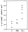
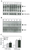
Interleukin-1 (IL-1), an inflammatory cytokine overexpressed in the neuritic plaques of Alzheimer's disease, activates astrocytes and enhances production and processing of beta-amyloid precursor protein (beta-APP). Activated astrocytes, overexpressing S100 beta, are a prominent feature of these neuritic plaques, and the neurite growth-promoting properties of S100 beta have been implicated in the formation of dystrophic neurites overexpressing beta-APP in neuritic plaques. These facts collectively suggest that elevated levels of the inflammatory cytokine IL-1 drive S100 beta and beta-APP overexpression and dystrophic neurite formation in Alzheimer's disease. To more directly assess this driver potential for IL-1, we analyzed IL-1 induction of S100 beta expression in vivo and in vitro, and of beta-APP expression in vivo. Synthetic IL-1 beta was injected into the right cerebral hemispheres of 13 rats. Nine additional rats were injected with phosphate-buffered saline, and seven rats served as uninjected controls. The number of astrocytes expressing detectable levels of S100 beta in tissue sections from IL-1-injected brains was 1.5 fold that of either control group (p < 0.01), while tissue S100 beta levels were approximately threefold that of controls (p < 0.05). The tissue levels of two beta-APP isoforms (approximately 130 and 135 kDa) were also significantly elevated in IL-1-injected brains (p < 0.05). C6 glioma cells, treated in vitro for 24 h with either IL-1 beta or IL-1 alpha, showed significant increases in both S100 beta and S100 beta mRNA levels. These results provide evidence that IL-1 upregulates both S100 beta and beta-APP expression, in vivo and vitro, and support the idea that overexpression of IL-1 in Alzheimer's disease drives astrocytic overexpression of S100 beta, favoring the growth of dystrophic neurites necessary for evolution of diffuse amyloid deposits into neuritic beta-amyloid plaques.
Figures


Similar articles
-
Correlation of astrocytic S100 beta expression with dystrophic neurites in amyloid plaques of Alzheimer's disease.J Neuropathol Exp Neurol. 1996 Mar;55(3):273-9. doi: 10.1097/00005072-199603000-00002. J Neuropathol Exp Neurol. 1996. PMID: 8786385 Free PMC article.
-
Interleukin-1 expression in different plaque types in Alzheimer's disease: significance in plaque evolution.J Neuropathol Exp Neurol. 1995 Mar;54(2):276-81. doi: 10.1097/00005072-199503000-00014. J Neuropathol Exp Neurol. 1995. PMID: 7876895
-
Microglial interleukin-1 alpha expression in brain regions in Alzheimer's disease: correlation with neuritic plaque distribution.Neuropathol Appl Neurobiol. 1995 Aug;21(4):290-301. doi: 10.1111/j.1365-2990.1995.tb01063.x. Neuropathol Appl Neurobiol. 1995. PMID: 7494597
-
Interleukin-1, neuroinflammation, and Alzheimer's disease.Neurobiol Aging. 2001 Nov-Dec;22(6):903-8. doi: 10.1016/s0197-4580(01)00287-1. Neurobiol Aging. 2001. PMID: 11754997 Review.
-
The role of activated astrocytes and of the neurotrophic cytokine S100B in the pathogenesis of Alzheimer's disease.Neurobiol Aging. 2001 Nov-Dec;22(6):915-22. doi: 10.1016/s0197-4580(01)00293-7. Neurobiol Aging. 2001. PMID: 11754999 Review.
Cited by
-
In vivo indomethacin treatment causes microglial activation in adult mice.Neurochem Res. 2000 Mar;25(3):357-62. doi: 10.1023/a:1007588903897. Neurochem Res. 2000. PMID: 10761979
-
Role of Activated Glia and of Glial Cytokines in Alzheimer's Disease: A Review.EOS. 1996;16(3-4):80-84. EOS. 1996. PMID: 24478534 Free PMC article.
-
Alzheimer Drug Trials: Combination of Safe and Efficacious Biologicals to Break the Amyloidosis-Neuroinflammation Vicious Cycle.ASN Neuro. 2020 Jan-Dec;12:1759091420918557. doi: 10.1177/1759091420918557. ASN Neuro. 2020. PMID: 32290675 Free PMC article. Review.
-
CCR5 deficiency accelerates lipopolysaccharide-induced astrogliosis, amyloid-beta deposit and impaired memory function.Oncotarget. 2016 Mar 15;7(11):11984-99. doi: 10.18632/oncotarget.7453. Oncotarget. 2016. PMID: 26910914 Free PMC article.
-
Does neuroinflammation fan the flame in neurodegenerative diseases?Mol Neurodegener. 2009 Nov 16;4:47. doi: 10.1186/1750-1326-4-47. Mol Neurodegener. 2009. PMID: 19917131 Free PMC article.
References
-
- Allore R, O’Hanlon D, Price R, Neilson K, Willard HF, Cox DR, Marks A, Dunn RJ. Gene encoding the β subunit of S100 protein is on chromosome 21: Implications for Down syndrome. Science. 1988;239:1311–1313. - PubMed
-
- Araki W, Kitaguchi N, Tokushima Y, Ishii K, Aratake H, Shimohama S, Nakamura S, Kimura J. Trophic effect of β-amyloid precursor protein on cerebral cortical neurons in culture. Biochem Biophys Res Commun. 1991;181:265–271. - PubMed
-
- Baggott PJ, Sheng JG, Cork L, Del Bigio MR, Brumback RA, Roberts GW, Mrak RE, Griffin WST. Expression of Alzheimer’s disease (AD)-related proteins during development in Down’s syndrome. Neurosci Soc Abstr. 1993;19:182.
-
- Ban EM, Sarlieve LL, Haour FG. Interleukin-1 binding sites on astrocytes. Neuroscience. 1993;52:725–733. - PubMed
-
- Bhattacharyya A, Oppenheim RW, Prevette D, Moore BW, Brackenbury X, Rattner N. S100 is present in developing chicken neurons and Schwann cells and promotes neuron survival in vivo. J Neurobiol. 1992;23:451–466. - PubMed
Publication types
MeSH terms
Substances
Grants and funding
LinkOut - more resources
Full Text Sources
Other Literature Sources
Medical

