Comparative Study
. 1996 Nov 12;93(23):12774-9.
doi: 10.1073/pnas.93.23.12774.

Novel dimeric interface and electrostatic recognition in bacterial Cu,Zn superoxide dismutase

Affiliations
Comparative Study

Novel dimeric interface and electrostatic recognition in bacterial Cu,Zn superoxide dismutase

Y Bourne et al. Proc Natl Acad Sci U S A. .

Abstract

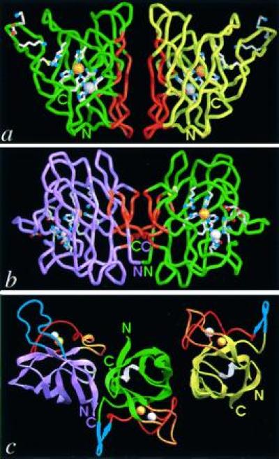
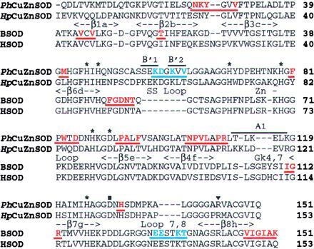
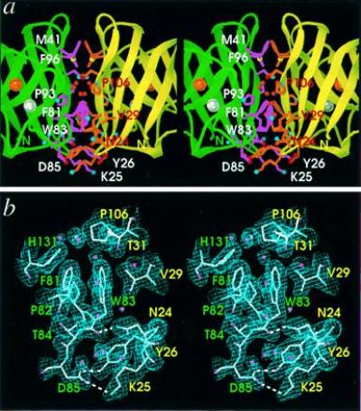
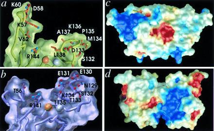
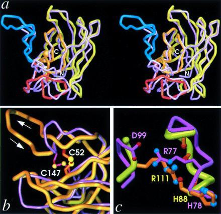
Eukaryotic Cu,Zn superoxide dismutases (CuZnSODs) are antioxidant enzymes remarkable for their unusually stable beta-barrel fold and dimer assembly, diffusion-limited catalysis, and electrostatic guidance of their free radical substrate. Point mutations of CuZnSOD cause the fatal human neurodegenerative disease amyotrophic lateral sclerosis. We determined and analyzed the first crystallographic structure (to our knowledge) for CuZnSOD from a prokaryote, Photobacterium leiognathi, a luminescent symbiont of Leiognathid fish. This structure, exemplifying prokaryotic CuZnSODs, shares the active-site ligand geometry and the topology of the Greek key beta-barrel common to the eukaryotic CuZnSODs. However, the beta-barrel elements recruited to form the dimer interface, the strategy used to forge the channel for electrostatic recognition of superoxide radical, and the connectivity of the intrasubunit disulfide bond in P. leiognathi CuZnSOD are discrete and strikingly dissimilar from those highly conserved in eukaryotic CuZnSODs. This new CuZnSOD structure broadens our understanding of structural features necessary and sufficient for CuZnSOD activity, highlights a hitherto unrecognized adaptability of the Greek key beta-barrel building block in evolution, and reveals that prokaryotic and eukaryotic enzymes diverged from one primordial CuZnSOD and then converged to distinct dimeric enzymes with electrostatic substrate guidance.


Figures

Figure 1
Figure 1
Comparison of the folds and dimer assemblies of P-class PhCuZnSOD with E-class bovine CuZnSOD (BSOD). PhCuZnSOD dimer Cα trace (green and yellow subunits) (a) and BSOD dimer (18) Cα trace (purple and green subunits) (b) viewed perpendicular to the dimer twofold axis and down into the active-site Cu for one subunit. Functionally important side chains for the active site, disulfide cysteines, and electrostatic recognition residues are displayed with white bonds and atom-colored spheres (red oxygen, blue nitrogen, and yellow sulfur atoms). Dimer interface regions in both P- and E-class CuZnSOD are highlighted (orange), and active-site metal ions are shown as spheres for the copper (orange) and zinc (silver). (c) PhCuZnSOD dimer (Right) and BSOD dimer (18) (Left) shown following superposition of one subunit (Cα atoms) from each. The central subunit (green) is displayed for PhCuZnSOD only and the subunit color code matches a and b. Loops are color-coded by function: blue for contributing to electrostatic attraction of substrate (loop 7,8 in BSOD, Upper Left, and SS loop in PhCuZnSOD, Upper Right), orange for forming the dimer interface (SS loop in BSOD and loop 7,8 in PhCuZnSOD), red for binding Zn ion, and white for the second Greek key connection across the end of the β-barrel. For loop nomenclature, see Fig. 2 legend. The view is down the dimer twofold axis and approximately 90° away from that in a and b. Figs. 1, 3a, 4, and 5 were generated with the Application Visualization System (AVS) (Advanced Visual Systems, Waltham, MA) and Figs. 1c and 3a were made with ribbons (19) implemented in AVS by Alexandre Shah.
Figure 2
Figure 2
Sequence and secondary structure of PhCuZnSOD compared with other P- and E-class CuZnSODs. Sequences of two bacterial CuZnSODs, PhCuZnSOD (21, 22), and HpCuZnSOD (Haemophilus parainfluenzae) (23), are aligned with sequences of eukaryotic bovine (BSOD, ref. 24) and human (HSOD, ref. 25) CuZnSODs, based on our structural comparison. Bacterial enzyme sequences share little homology with eukaryotic sequences [e.g., 28% identity between PhCuZnSOD and BSOD (10)] but display mutual sequence similarities of about 70% and share a common pattern of insertions/deletions (2, 23). For both the PhCuZnSOD and BSOD sequences, residues in the dimer interface are color-coded and underlined in orange, and those involved in the long-range electrostatic attraction of the substrate anion are in blue. The common CuZnSOD secondary structural elements are represented between the two pairs of sequences, with bounds for PhCuZnSOD. β-strands are named by letter in sequence order and by number clockwise around the β-barrel (18), and those specific to PhCuZnSOD are shown above the sequence. Cu and Zn ligands are marked with an asterisk, the buried Asp with ▪, and the catalytically important Arg with ▾. Fig. 2 was generated with framemaker (Frame Technology Corporation, San Jose, CA).
Figure 3
Figure 3
PhCuZnSOD dimer interface structure and electron density. Stereo pairs are oriented with the twofold axis vertical to match Fig. 1a. Labels indicate one-letter amino acid code and residue number. (a) Close-up displaying key side chains of the dimer interface with light purple bonds and white labels (for green subunit) or orange bonds and red labels (for yellow subunit) and atom-colored spheres (see Fig. 1a). The Asn-105 side chain and main-chain atoms of Ala-94, Asn-105, Pro-106, and Leu-108 hydrogen bond with the internal ring of buried water molecules, shown as red spheres. Active-site metal ions are shown as spheres for the copper (orange) and zinc (silver). (b) Refined model (atom-colored), solvent water molecules (pink spheres), and 2FoFc electron density map (contoured at 1.0 SD above the mean) at the dimer interface. Hydrogen bonds spanning the interface are shown as white dashed lines and residue labels are color-coded by subunit. b was generated with turbo-frodo (15).
Figure 4
Figure 4
Conservation and variation of the active channel shape and electrostatic potential between P- and E-class CuZnSODs. (a) Active-site channel cross-section for P-class PhCuZnSOD and (b) E-class BSOD. Whereas the shape and dimensions are conserved between the two classes (at left, PhCuZnSOD Val-62 coincides with BSOD Thr-56 in the SS loop, and at right, PhCuZnSOD Leu-138 coincides with BSOD Thr-135 in loop 7,8), the structural location, conformation, and identity of the residues producing the long-range electrostatic attraction of the substrate anion are dramatically different, suggesting separate divergent evolution followed by convergence. Electrostatic potential mapped onto the molecular surface of the PhCuZnSOD dimer (c) (oriented to match Fig. 1a) and the BSOD dimer (d) (oriented to match Fig. 1b) and colored as blue positive potential (>3kT/q) and red negative potential (<−3kT/q). The network of charged residues Lys-57, Asp-58, and Lys-60, in the extended PhCuZnSOD SS loop (upper left) forms the positive potential for substrate attraction, whereas Glu-131, Glu-132, and Lys-134 in the extended loop 7,8 (lower right) provide the equivalent function in BSOD.
Figure 5
Figure 5
β-Barrel rearrangements. (a) Stereo pair superposition of the Cα traces of PhCuZnSOD (yellow) and BSOD (purple) subunits viewed perpendicular to the β-barrel axis (approximately 90° from Fig. 1a), showing the large insertion in PhCuZnSOD SS loop (blue, left) as well as distortions in the bottom β-sheet (center and right). The functional elements and metal ions of PhCuZnSOD are color-coded as in Fig. 1c. (b) Close-up view of SS loop in superimposed subunits, as for a. PhCuZnSOD disulfide (orange bonds with yellow spheres) Cys-52 (aligned beneath BSOD Asn-51) is located on the other side of the SS loop relative to BSOD disulfide (magenta bonds with yellow spheres) Cys-55 (both at top), while the position of PhCuZnSOD Cys-147 (BSOD Cys-144) is conserved (both below). (c) Close-up view of Zn loop in superimposed subunits, as for a, showing the functional equivalence of PhCuZnSOD Arg-111 (orange bonds and atom-colored spheres) and BSOD Arg-77 (light purple bonds and atom-colored spheres) in stabilizing the Zn loop. BSOD Arg-77 forms a salt bridge with Asp-99 (left), while PhCuZnSOD Arg-111 hydrogen bonds with four main-chain carbonyl oxygen atoms (right).

References

    1. Beyer W, Imlay J, Fridovich I. Prog Nucleic Acid Res Mol Biol. 1991;40:221–253. - PubMed
    1. Bordo D, Djinovic K, Bolognesi M. J Mol Biol. 1994;238:366–386. - PubMed
    1. Grace S C. Life Sci. 1990;47:1875–1886. - PubMed
    1. Kroll J S, Langford P R, Wilks K E, Keil A D. Microbiology. 1995;141:2271–2279. - PubMed
    1. St. John G, Steinman H M. J Bacteriol. 1996;178:1578–1584. - PMC - PubMed

Publication types

MeSH terms

LinkOut - more resources