

Superoxide-mediated clastogenesis and anticlastogenic effects of exogenous superoxide dismutase
- PMID: 8917499
- PMCID: PMC24000
- DOI: 10.1073/pnas.93.23.12799
Superoxide-mediated clastogenesis and anticlastogenic effects of exogenous superoxide dismutase
Abstract
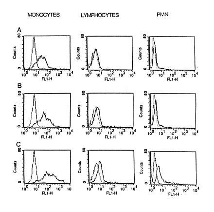
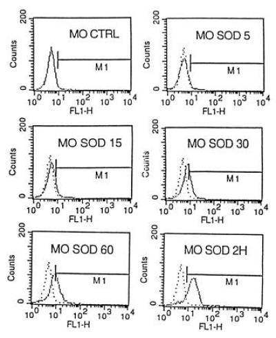
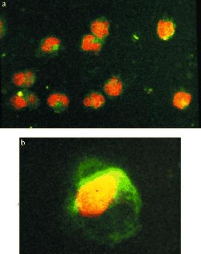
Superoxide-mediated clastogenesis is characteristic for various chronic inflammatory diseases with autoimmune reactions and probably plays a role in radiation-induced clastogenesis and in the congenital breakage syndromes. It is consistently prevented by exogenous superoxide dismutase (SOD), but not by heat-inactivated SOD, indicating that the anticlastogenic effect is related to the catalytic function of the enzyme. Increased superoxide production by activated monocytes/macrophages is followed by release of more long-lived metabolites, so-called clastogenic factors, which contain lipid peroxidation products, unusual nucleotides of inosine, and cytokines such as tumor necrosis factor alpha. Since these components are not only clastogenic, but can stimulate further superoxide production by monocytes and neutrophils, the genotoxic effects are self-sustaining. It is shown here that anticlastogenic effects of exogenous SOD are preserved despite extensive washing of the cells and removal of all extracellular SOD. Using flow cytometry and confocal laser microscopy, rapid adherence of the fluorescently labeled enzyme to the cell surface could be observed with slow uptake into the cell during the following hours. The degree of labeling was concentration and time dependent. It was most important for monocytes, compared with lymphocytes, neutrophils, and fibroblasts. The cytochrome c assay showed significantly diminished O2- production by monocytes, pretreated with SOD and washed thereafter. The preferential and rapid binding of SOD to monocytes may be of importance not only for the superoxide-mediated genotoxic effects, described above, but also from a therapeutic standpoint. It can explain the observation that beneficial effects of injected SOD lasted for weeks and months despite rapid clearance of the enzyme from the blood stream according to pharmacodynamic studies.
Figures
References
MeSH terms
Substances
LinkOut - more resources
Full Text Sources
