. 1996 Nov 12;93(23):12840-4.
doi: 10.1073/pnas.93.23.12840.

Repair of thalassemic human beta-globin mRNA in mammalian cells by antisense oligonucleotides

Affiliations

Repair of thalassemic human beta-globin mRNA in mammalian cells by antisense oligonucleotides

H Sierakowska et al. Proc Natl Acad Sci U S A. .

Abstract

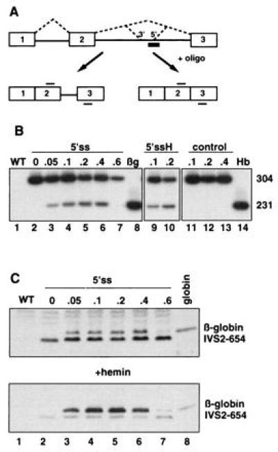
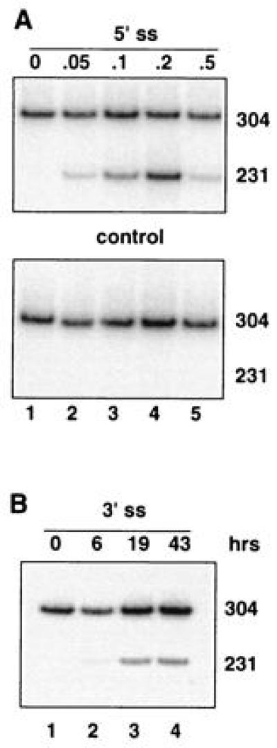
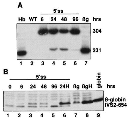
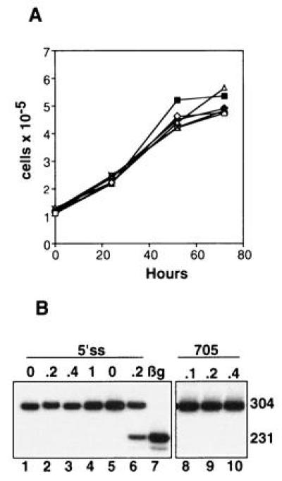
In one form of beta-thalassemia, a genetic blood disorder, a mutation in intron 2 of the beta-globin gene (IVS2-654) causes aberrant splicing of beta-globin pre-mRNA and, consequently, beta-globin deficiency. Treatment of mammalian cells stably expressing the IVS2-654 human beta-globin gene with antisense oligonucleotides targeted at the aberrant splice sites restored correct splicing in a dose-dependent fashion, generating correct human beta-globin mRNA and polypeptide. Both products persisted for up to 72 hr posttreatment. The oligonucleotides modified splicing by a true antisense mechanism without overt unspecific effects on cell growth and splicing of other pre-mRNAs. This novel approach in which antisense oligonucleotides are used to restore rather than to down-regulate the activity of the target gene is applicable to other splicing mutants and is of potential clinical interest.


Figures

Figure 1
Figure 1
(A) Splicing of human β-globin IVS2-654 pre-mRNA in the presence of an antisense oligonucleotide. Boxes, exons; solid lines, introns; dashed lines, both correct and aberrant splicing pathways; thick bar, oligonucleotide antisense to the aberrant 5′ splice site; thin bars above and below exon sequences, primers used in the RT-PCR reaction. The aberrant 5′ splice site created by IVS2-654 mutation and the cryptic 3′ splice site activated upstream are indicated. (B) Correction of splicing of IVS2-654 pre-mRNA in HeLa cells by antisense oligonucleotide targeted to the aberrant 5′ splice site (5′ss). Analysis of total RNA by RT-PCR. Lanes: 1, wild-type (WT) HeLa cells; 8, HeLa cell line expressing normal human β-globin (βg); 14, RNA from human blood (Hb); 2–7, IVS2-654 HeLa cells treated with increasing concentrations of the oligonucleotide (indicated in micromoles at the top); 9 and 10, IVS2-654 HeLa cells treated with oligonucleotide followed by hemin (H) (16); 11–13, IVS2-654 HeLa cells treated with increasing concentrations of the scrambled oligonucleotide. The numbers on the right indicate the size, in nucleotides, of the RT-PCR products representing the aberrantly (304) and correctly (231) spliced RNAs. (C) Restoration of β-globin expression by 5′ss oligonucleotide in IVS2-654 HeLa cells. Immunoblot of total protein with anti-human hemoglobin antibody. Concentration of the oligonucleotide in micromoles is indicated at the top (lanes 2–7); in lane 8, human globin (Sigma) was used as a marker. (Lower) Cells were treated with hemin preceding the isolation of proteins. The positions of human β-globin and the prematurely terminated β-globin IVS2-654 polypeptide are indicated. Time of exposure of the autoradiogram in Lower was 1/5th of that of the Upper.
Figure 3
Figure 3
Dose- and time-dependent correction of splicing in oligonucleotide-treated IVS2-654 NIH 3T3 cells. RT-PCR assay. (A) Cells treated with increasing concentrations of the oligonucleotide targeted to the aberrant 5′ splice site (Upper) or of the control, scrambled oligonucleotide (Lower). (B) Time course of the correction of splicing after termination of treatment with 0.2 μM oligonucleotide targeted to the cryptic 3′ splice site activated by the IVS2-654 mutation. All designations are as in Figs. 1 and 2.
Figure 2
Figure 2
Time course of restoration of correct splicing and β-globin expression in HeLa IVS2-654 cells by 0.2 μM 5′ss oligonucleotide. (A) RT-PCR assay. (B) Immunoblot. Time after termination of oligonucleotide treatment is indicated at the top. H, hemin treatment of the cells. All other designations are as in Fig. 1.
Figure 4
Figure 4
Specificity of oligonucleotide treatments. (A) Lack of effect of oligonucleotides on cell growth. HeLa IVS2-654 cells were treated with Lipofectamine–oligonucleotide complexes as described. Cells were counted at the end of the 10 hr treatment (0 hr) and at 24, 52, and 72 hr thereafter. Each point on the curve represents the average of duplicate counts of two independently treated samples; the observed differences are within experimental error (one SD). □, Lipofectamine alone. The remaining samples were treated with Lipofectamine complexed with the following 0.2 μM oligoribonucleotides: 2′-O-methyl phosphorothioates, ⋄, 5′ss, ×, 3′ss, ♦, scrambled; 2′-O-methyl phosphodiesters, ▵, 5′ss, ▪, 3′ss. (B) Lack of effect of control oligonucleotides. Treatment of HeLa IVS2-654 consensus cell line (lanes 1–4, see text) or HeLa IVS2-654 cell line (lanes 5 and 6, as positive control) with 5′ss 2′-O-methyl phosphorothioate oligoribonucleotide. Lanes 8–10, treatment of HeLa IVS2-654 cells with oligonucleotide 705 targeted 44 nucleotides downstream from the aberrant 5′ splice site (see text). The RT-PCR assay and all designations are as described in the legend to Fig. 1B. Lane 7, HeLa cell line expressing normal human β-globin.

References

    1. Schwartz E, Benz E J. In: Hematology, Basic Principles and Practice. Hoffman R, Benz E J, Shattil S J, Furie B, Cohen H J, editors. New York: Churchill Livingstone; 1995. pp. 586–610.
    1. Huisman T H J. Br J Haematol. 1990;75:454–457. - PubMed
    1. Cheng T-C, Orkin S H, Antonorakis S E, Potter M J, Sexton J P, Giardina P J V, Li A, Kazazian H H. Proc Natl Acad Sci USA. 1984;81:2821–2826. - PMC - PubMed
    1. Huang S-Z, Zeng F-Y, Ren Z-R, Lu Z-H, Rodgers G P, Schechter A N, Zeng Y-T. Br J Haematol. 1994;88:541–546. - PubMed
    1. Busslinger M N, Moschonas N, Flavell R A. Cell. 1981;27:289–296. - PubMed

Publication types

MeSH terms

LinkOut - more resources