. 1996 Nov 12;93(23):12890-5.
doi: 10.1073/pnas.93.23.12890.

Development of pilus organelle subassemblies in vitro depends on chaperone uncapping of a beta zipper

Affiliations

Development of pilus organelle subassemblies in vitro depends on chaperone uncapping of a beta zipper

E Bullitt et al. Proc Natl Acad Sci U S A. .

Abstract

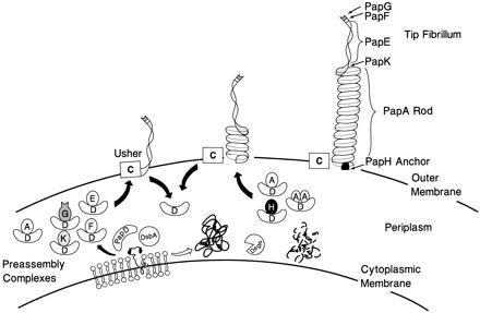
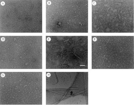
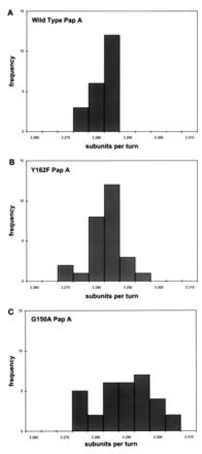
The major subassemblies of virulence-associated P pili, the pilus rod (comprised of PapA) and tip fibrillum (comprised of PapE), were reconstituted from purified chaperone-subunit complexes in vitro. Subunits are held in assembly-competent conformations in chaperone-subunit complexes prior to their assembly into mature pili. The PapD chaperone binds, in part, to a conserved motif present at the C terminus of the subunits via a beta zippering interaction. Amino acid residues in this conserved motif were also found to be essential for subunit-subunit interactions necessary for the formation of pili, thus revealing a molecular mechanism whereby the PapD chaperone may prevent premature subunit-subunit interactions in the periplasm. Uncapping of the chaperone-protected C terminus of PapA and PapE was mimicked in vitro by freeze-thaw techniques and resulted in the formation of pilus rods and tip fibrillae, respectively. A mutation in the leading edge of the beta zipper of PapA produces pilus rods with an altered helical symmetry and azimuthal disorder. This change in the number of subunits per turn of the helix most likely reflects involvement of the leading edge of the beta zipper in forming a right-handed helical cylinder. Organelle development is a fundamental process in all living cells, and these studies shed new light on how immunoglobulin-like chaperones govern the formation of virulence-associated organelles in pathogenic bacteria.


Figures

Figure 1
Figure 1
Model of pilus assembly. Details of the model are discussed in the text. PapD has an immunoglobulin-like three-dimensional structure (9) and is the prototype member of a large family of PapD-like chaperones required for pilus assembly in Gram-negative bacteria (13). DsbA mediates disulfide bond formation in PapD and in the pilus subunits (14). DsbA is required for the correct folding of PapD. Closed arrow depicts pathway subunits travel in the presence of PapD chaperone. Subunits misfold and are proteolytically degraded by the DegP protease in the absence of an interaction with PapD (open arrow) (C.H.J. and S.J.H., unpublished work). Chaperone–subunit complexes are targeted to the PapC usher protein (12) where pilus assembly occurs; an essential step in assembly is the displacement of PapD (uncapping). The PapD chaperone has been shown to interact with pilus subunits, in part, via the COOH terminal motif (15). Binding of PapD to an assembly surface on the subunits modulates the formation of the pilus rod.
Figure 2
Figure 2
In vitro assembly of pilus-like or tip fibrillum-like fibers after chaperone uncapping. Panels A–H are electron micrographs of chaperone–subunit complexes following rapid freeze-thaw conditions. PapD–PapA complexes (a mixture of DA, 1:1 PapD–PapA, and DA2, 1:2 PapD–PapA) before (A) and after three (B) or 10 (C) freeze-thaw cycles. (D and E) The PapD–PapA complexes after 10 freeze-thaw cycles in the presence (D) or absence (E) of a 10-fold excess of free PapD. Freeze-thaw treatment of purified PapD-PapE complex and PapD-PapK complex is shown in F and G, respectively. H is an electron micrograph of purified P pili showing the composite architecture of the fiber: a tip fibrillum (→) joined end to end to a pilus rod. Magnification bar in E = 500 Å.
Figure 3
Figure 3
Point mutations in the Beta zipper differentially effect chaperone–subunit complex formation and subunit–subunit interactions. (A) Isoelectric focusing gel electrophoresis demonstrating DA (1:1) and DA2 (1:2) complexes. Note that substitution of Tyr-162 diminishes (Y162F, lane 3) or abolishes (Y162L, lane 4) the formation of the DA complex while having no effect on the DA2 complex. In contrast the G150T substitution greatly decreases the DA2 band while having no effect on the DA complex (lane 7). Note that the samples in lanes 1–5 were loaded from the top comb position while the samples in lanes 6 and 7 were loaded from the bottom comb position, accounting for a somewhat cleaner gel. (B) Subunit–subunit interactions are stable at 25°C in 1.5% SDS, however the interactions are disrupted by heating the samples to 95°C (8). Periplasmic extracts were run on SDS/PAGE after treatment at 25°C (odd lanes) or 95°C (even lanes). The PapA subunit complexes were visualized by Western blot analysis. Note that with wild-type PapA coexpressed with PapD, subunit complexes migrating with an apparent mobility of dimers, trimers, and tetramers are resolved at 25°C (lane 3). The Tyr-162 substitution to leucine blocks the formation of PapA multimers (lane 11) while the Gly-150 substitution to threonine seems to favor stable trimer formation (lane 17).
Figure 4
Figure 4
G150A-PapA subunit alters the helical symmetry of the Pap pilus rod. The helical symmetry of pili purified from the bacterial cell surface containing wild-type (A), Y162F (B), and (C) G150A PapA was calculated as described (19). The symmetry of the Y162F pili is unchanged from wild type: 3.281 ± 0.006 with a helical pitch of 24.77 ± 0.87 Å (n = 27) vs. 3.279 ± 0.003 u/t with a helical pitch of 24.87 ± 0.41 Å (n = 21), respectively. The symmetry of the G150A pili is more variable and has a tighter twist, 3.287 ± 0.009 u/t and a helical pitch of 24.67 ± 1.18 Å (n = 32); i.e., this mutant has a larger standard deviation and more u/t of the helix than wild-type pili.

References

    1. Hultgren S J, Abraham S N, Caparon M G, Falk P, St. Geme J W, III, Normark S. Cell. 1993;73:887–901. - PubMed
    1. Hultgren S J, Normark S, Abraham S N. Annu Rev Microbiol. 1991;45:383–415. - PubMed
    1. Kuehn M J, Heuser J, Normark S, Hultgren S J. Nature (London) 1992;356:252–255. - PubMed
    1. Gong M, Makowski L. J Mol Biol. 1992;228:735–742. - PubMed
    1. Bullitt E, Makowski L. Nature (London) 1995;373:164–167. - PubMed

Publication types

MeSH terms

LinkOut - more resources