. 1996 Nov 12;93(23):12896-901.
doi: 10.1073/pnas.93.23.12896.

Reconstitution of human replication factor C from its five subunits in baculovirus-infected insect cells

Affiliations

Reconstitution of human replication factor C from its five subunits in baculovirus-infected insect cells

J Cai et al. Proc Natl Acad Sci U S A. .

Abstract

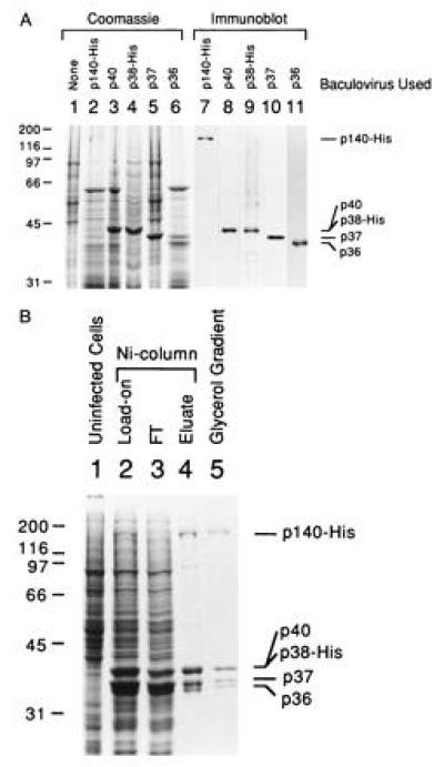
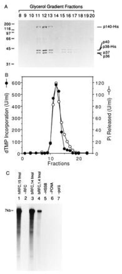
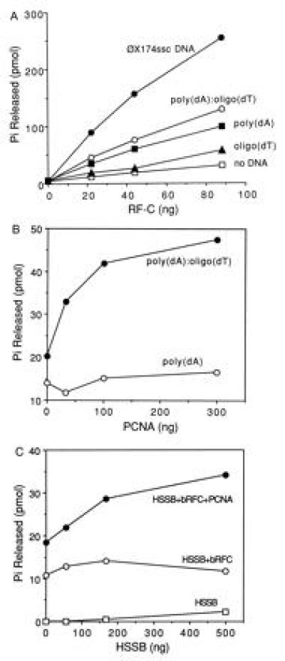
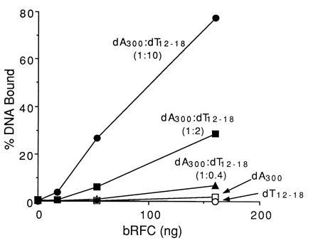
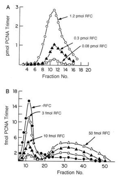
Human replication factor C (RFC, also called activator 1) is a five-subunit protein complex (p140, p40, p38, p37, and p36) required for proliferating cell nuclear antigen (PCNA)-dependent processive DNA synthesis catalyzed by DNA polymerase delta or epsilon. Here we report the reconstitution of the RFC complex from its five subunits simultaneously overexpressed in baculovirus-infected insect cells. The purified baculovirus-produced RFC appears to contain equimolar levels of each subunit and was shown to be functionally identical to its native counterpart in (i) supporting DNA polymerase delta-catalyzed PCNA-dependent DNA chain elongation; (ii) catalyzing DNA-dependent ATP hydrolysis that was stimulated by PCNA and human single-stranded DNA binding protein; (iii) binding preferentially to DNA primer ends; and (iv) catalytically loading PCNA onto singly nicked circular DNA and catalytically removing PCNA from these DNA molecules.


Figures

Figure 1
Figure 1
Reconstitution of RFC from its five subunits in baculovirus-infected HF cells. (A) Overexpression of the RFC subunits in baculovirus-infected HF cells. The five subunits of RFC were individually expressed in HF cells and cell lysates were analyzed by SDS/9% PAGE followed by staining with Coomassie brilliant blue (lanes 1–6) or by Western blot analysis (lanes 7–11). The additions to each lane were: lane 1, 30 μg of uninfected HF cell extract; lanes 2–6, 30 μg of extract from cells producing the p140, p40, p38, p37, and p36 subunit, respectively; lanes 7–11, immunoblots of cell lysates as described in lanes 2–6. Each blot was probed with specific antibodies against the corresponding subunit as follows: lane 7, p140; lane 8, p40; lane 9, p38; lane 10, p37; lane 11, p36. Molecular weight markers are indicated at the left of the figure and the position of each of the RFC subunits are indicated at the right. (B) Purification of reconstituted bRFC. The bRFC was reconstituted by coinfecting HF cells with all five recombinant viruses capable of expressing each RFC subunit. The reconstituted RFC was purified from nuclear extracts of the coinfected cells by Ni-affinity chromatography and subsequent glycerol gradient centrifugation as described. Proteins were visualized after SDS/PAGE by Coomassie blue staining. The additions to each lane were: lane 1, 30 μg of uninfected HF cell extract; lane 2, 25 μg of infected cell nuclear extract (Ni column load on); lane 3, 25 μg of protein that flowed through the Ni column; lane 4, 1.4 μg of Ni column eluate; lane 5, 0.9 μg of protein from glycerol gradient fractions 11–13. The markers at the left and right of the figure are as indicated in A.
Figure 2
Figure 2
Cosedimentation of RFC-dependent DNA synthesis activity and ATPase activity through a 15–35% glycerol gradient. (A) SDS/PAGE analysis. The bRFC, eluted from a Ni-column (Fig. 1B), was further purified by two consecutive 15–35% glycerol gradient centrifugations. Following acetone precipitation and centrifugation, the pellets were analyzed by SDS/PAGE followed by Coomassie staining. The numbers at the top of the figure represent the fraction analyzed. The bands that migrated between 55–70 kDa in all lanes were artifactual. (B) RFC-dependent DNA synthesis and DNA-dependent ATPase activity. Each glycerol gradient fraction (0.5 μl after 3-fold dilution) was assayed for its ability to support DNA synthesis in the presence of a multiply primed poly(dA)4500/oligo (dT)12–18 template and ATPase activity as described. (C) RFC-dependent nucleotide incorporation using a singly primed M13 DNA template. Reactions were carried out as described using 4.4 fmol of M13 DNA template prior to separation through alkaline agarose gel followed by autoradiography. Reactions shown in lanes 1 and 2 were carried out in the presence and absence of 15 fmol of hRFC, respectively; reactions shown in lanes 3–7 were carried out with bRFC as follows: lane 3, 14 fmol of bRFC; lane 4, 1.4 fmol of bRFC; lanes 5–7, 14 fmol of bRFC in the absence or presence of HSSB, PCNA, or pol δ as indicated. Nucleotide incorporation (pmol), measured following acid precipitation and liquid scintillation counting, was as follows: lane 1, 26; lane 2, 0.4; lane 3, 24.4; lane 4, 8.8; lane 5, 2.8; lane 6, 0.48; lane 7, 0.08.
Figure 3
Figure 3
Characterization of bRFC ATPase activity. (A) Stimulation of bRFC ATPase by DNA effectors. ATPase reactions were carried out as described. Reaction mixtures contained bRFC in amounts as indicated, in the absence or presence of 12.5 μM (nucleotide concentration) of the following DNAs: oligo(dT)12–18, poly(dA)4500, poly(dA)4500/oligo(dT)12–18 (20:1 nucleotide ratio), or φX174 circular ssDNA. (B) PCNA stimulation of RFC ATPase activity. PCNA was added in amounts as indicated to reaction mixtures containing 15 ng of bRFC in the presence of 12.5 μM of poly(dA)4500 or poly(dA)4500/oligo(dT)12–18. (C) HSSB stimulation of RFC ATPase activity. HSSB was added in amounts as indicated to reaction mixtures containing poly(dA)4500/oligo(dT)12–18 in the absence or presence of 10 ng of bRFC and/or 40 ng of PCNA.
Figure 4
Figure 4
bRFC preferentially binds to DNA primer ends. Binding of bRFC to DNA was examined using a nitrocellulose filter binding assay as described. bRFC, in amounts as indicated, was incubated with poly(dA)300, oligo(dT)12–18, or poly(dA)300/oligo(dT)12–18 at a molar ratio of 1:0.4, 1:2 and 1:10, respectively, as indicated. The 100% value represented 40 fmol of input DNA.
Figure 5
Figure 5
bRFC loads and unloads PCNA onto and off DNA. (A) bRFC catalyzed loading of PCNA onto singly nicked pBluescript DNA. Reaction mixtures (100 μl) containing 40 mM Tris·HCl (pH 7.5), 0.5 mM DTT, 20 μg/ml BSA, 7 mM Mg (OAc)2, 2 mM ATP, 0.5 pmol of singly nicked pBluescript plasmid DNA, 5.2 pmol of 32P-labeled PCNA trimer (574 cpm/fmol) and bRFC (as indicated), were incubated for 10 min at 37°C. Each reaction mixture was then filtered through a 6 ml Bio-Gel A15m column equilibrated with buffer containing 0.02 M Tris·HCl (pH 7.5), 0.1 mM EDTA, 40 μg/ml bovine serum albumin, 8 mM MgCl2, 4% glycerol, 5 mM DTT, and 0.1 M NaCl at 4°C. Fractions (180 μl) were collected and subjected to Cerenkov counting to detect the elution of the labeled PCNA. The peak shown in the graph represented PCNA that eluted in the excluded volume, i.e., PCNA complexed with DNA. (B) bRFC unloads 32P-PCNA catalytically from singly nicked DNA. 32P-PCNA was assembled onto DNA as described in A and the product isolated by BioGel A15m separation. The isolated PCNA–DNA complexes were then incubated in the presence or absence of bRFC (amounts as indicated) in reaction mixtures as described in A prior to gel filtration to resolve free 32P-PCNA from the 32P-PCNA–DNA complex. Both included and excluded material were subjected to Cerenkov counting as described above.

References

    1. Lee S-H, Eki T, Hurwitz J. Proc Natl Acad Sci USA. 1989;86:7361–7365. - PMC - PubMed
    1. Tsurimoto T, Stillman B. Mol Cell Biol. 1989;9:609–619. - PMC - PubMed
    1. Lee S-H, Kwong A D, Pan Z-Q, Hurwitz J. J Biol Chem. 1991;266:594–602. - PubMed
    1. Tsurimoto T, Stillman B. Proc Natl Acad Sci USA. 1990;87:1023–1027. - PMC - PubMed
    1. Yoder B L, Burgers P M. J Biol Chem. 1991;266:22689–22697. - PubMed

Publication types

MeSH terms

LinkOut - more resources