. 1996 Nov 12;93(23):12902-7.
doi: 10.1073/pnas.93.23.12902.

Direct physical interaction between DnaG primase and DnaB helicase of Escherichia coli is necessary for optimal synthesis of primer RNA

Affiliations

Direct physical interaction between DnaG primase and DnaB helicase of Escherichia coli is necessary for optimal synthesis of primer RNA

Y B Lu et al. Proc Natl Acad Sci U S A. .

Abstract

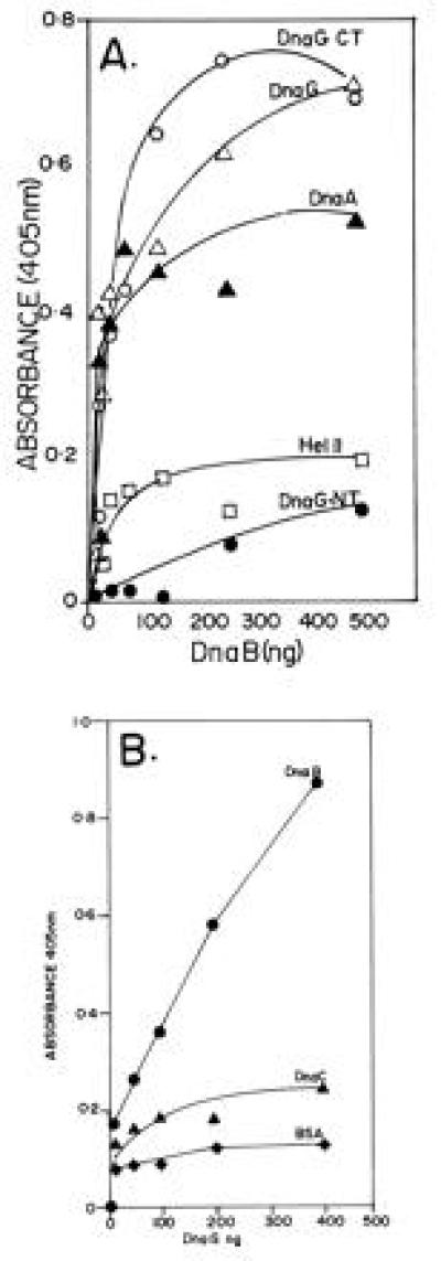
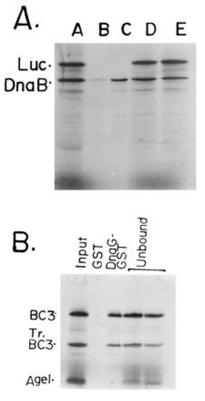
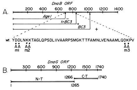
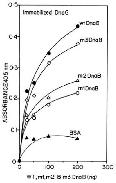
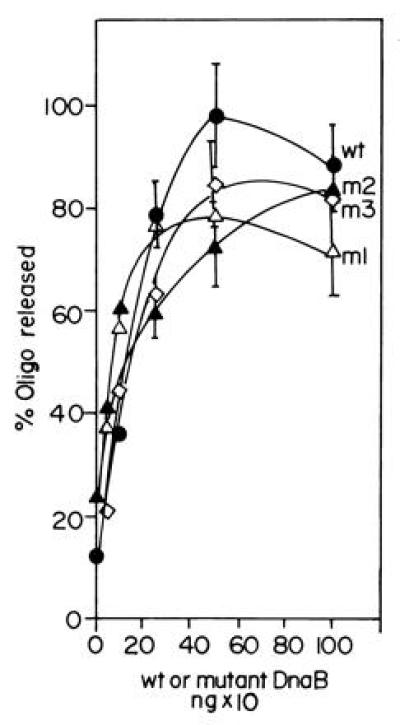
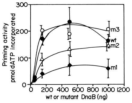
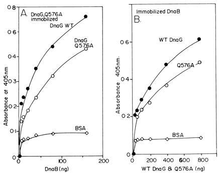
The primase DnaG of Escherichia coli requires the participation of the replicative helicase DnaB for optimal synthesis of primer RNA for lagging strand replication. However, previous studies had not determined whether the activation of the primase or its loading on the template was accomplished by a helicase-mediated structural alteration of the single-stranded DNA or by a direct physical interaction between the DnaB and the DnaG proteins. In this paper we present evidence supporting direct interaction between the two proteins. We have mapped the surfaces of interaction on both DnaG and DnaB and show further that mutations that reduce the physical interation also cause a significant reduction in primer synthesis. Thus, the physical interaction reported here appears to be physiologically significant.


Figures

Figure 1
Figure 1
ELISA showing the interaction of DnaG primase with DnaB helicase. (A) Interaction between immobilized DnaG with DnaB in solution. Full-length DnaG and its N-terminal (DnaG-NT) and C-terminal (DnaG-CT) peptides, DnaA, and helicase II (Hel II) were immobilized on plastic surfaces of the wells of microtiter plates and were challenged with various amounts of DnaB in solution. Note that full-length DnaG and its C-terminal peptide show clear binding to DnaB. The DnaA protein, used as a positive control also binds, as expected, to DnaB. In contrast the N-terminal peptide of DnaG and helicase II (negative control) elicited low or background levels of binding signal. (B) Reciprocal binding of immobilized DnaB to DnaG in solution. DnaB, DnaC (negative control), and BSA (negative control) were immobilized on the plastic surface of microtiter plates and challenged with various amounts of DnaG in solution. Whereas DnaG readily bound to immobilized DnaB, there was only low levels of binding of DnaG to immobilized DnaC and BSA.
Figure 2
Figure 2
GST-affinity chromatography showing the binding of full-length DnaB and its peptides with DnaG-GST fusion protein affinity matrix. (A) Full-length DnaB and luciferase (Luc; negative control) were labeled with 35S-methionine by coupled in vitro transcription-translation in rabbit reticulocyte extracts. An approximately equimolar mixture of the two labeled proteins were loaded on to the control GST-glutathione-agarose and the DnaG-GST-glutathione-agarose matrices. The flow through fraction was collected and analyzed. The beads were washed and the proteins eluted and resolved by SDS/PAGE. An autoradiogram of the gel shows: lane 1, input mixture of full-length DnaB, a truncated product of DnaB (N-terminal fragment) and full-length luciferase (Luc); lane 2, protein bound to the control GST-agarose matrix; lane 3, protein bound to the DnaG-GST-agarose matrix. Note that full-length DnaB, its truncated peptide, that was generated during the labeling process, were selectively retained on the DnaG-GST matrix but not on the control GST-matrix. No luciferase bound to the control or to the DnaG-GST matrices. (B) DNA template encoding the BC3 and AgeI (see Fig. 3) peptides of DnaB were labeled in vitro as described. The In vitro labeling process generated the tr.BC3 (Tr.BC3) peptide. The truncation must have generated a N-terminal peptide because of the upstream location of the T7 promoter used to transcribe the DNA. Lanes: 1, input mixture of BC3, TrBC3 and the AgeI peptides; 2, binding to the control GST matrix; 3, binding to the DnaG-GST matrix; 4, flow through from the GST matrix; 5, flow through from the DnaG-GST matrix. Note that the BC3 and tr.BC3 peptides (≈40–50% of the input) were retained on the DnaG-GST but not on the control GST matrix. The AgeI peptide was not retained by either matrices and was recovered in the flow through fractions.
Figure 3
Figure 3
Maps showing the interaction surfaces that participated in the DnaB–DnaG interaction. (A) Map showing DnaB sequence encoding the AgeI, tr.BC3, and BC3 peptides. The region between the AgeI and tr.BC3 binds to DnaB. The amino acid sequence showing the locations of the m1, m2, and m3 mutants that were used to further narrow down the interaction region. (B) The sequence encoding DnaG showing the locations of 1265-bp-long DNA encoding the N-terminal peptide that did not bind to DnaB and the 475-bp-long sequence encoding the C-terminal peptide that bound to DnaB. The numbers refer to base pair coordinates.
Figure 4
Figure 4
ELISA showing the binding of the m1, m2, and m3 mutant forms of DnaB with immobilized DnaG protein. The data are the averages of three separate experiments. The extent of binding is in the following decreasing order: wt DnaB > m3 > m2 > m1. BSA was used as a negative control.
Figure 5
Figure 5
Helicase assay showing the activities of wt and the m1, m2, and m3 forms of DnaB. The mutant forms of DnaB helicase have activities in the range of at least 85–95% of that of the wt enzyme. The helicase assays were performed by hybridizing a labeled 17-mer oligonucleotide to single-stranded M13 DNA and measuring the ATP-dependent unwinding by nondenaturing PAGE. The released radioactive oligodeoxynucleotide bands were excised from the gel and counted.
Figure 6
Figure 6
The relative abilities of wt and the mutant forms of DnaB to activate the ability of wt DnaG to synthesize RNA primers on uncoated M13 ssDNA. Note that the m1 mutant shows a 4- to 5-fold reduction in its ability to promote primer synthesis in collaboration with wt DnaG primase. The assay for primer synthesis is described in the text. The m3 mutant does not show detectable reduction in primer synthesis as compared the wt DnaB where as m2 shows more activity than m1 but less than that of the wt helicase.
Figure 7
Figure 7
ELISA showing the relative interactions of DnaB with wt DnaG and the mutant form Q576A. (A) Interaction of immobilized wt DnaG and Q576A with wt DnaB in solution. A typical set of results is shown. The wt DnaG consistently bound better than the Q576A protein DnaB in solution. (B) The experiment was performed in converse by immobilizing a constant amount (1 μg per well) of normal DnaB protein and challenging it with equivalent amount of wt and the Q576A protein. The wt DnaG, now present in solution, consistently bound more strongly to the immobilized DnaG than the mutant form.

References

    1. Alberts B M. Trends Genet. 1985;1:26–30.
    1. Rowen L, Kornberg A. J Biol Chem. 1978;253:758–764. - PubMed
    1. Arai K-I, Kornberg A. Proc Natl Acad Sci USA. 1979;76:4308–4313. - PMC - PubMed
    1. Kelly T J. J Biol Chem. 1988;263:17889–17892. - PubMed
    1. LeBowitz J, McMacken R. J Biol Chem. 1986;261:4738–4748. - PubMed

Publication types

MeSH terms

LinkOut - more resources