. 1996 Nov 12;93(23):13066-71.
doi: 10.1073/pnas.93.23.13066.

Identification and characterization of differentially expressed cDNAs of the vector mosquito, Anopheles gambiae

Affiliations

Identification and characterization of differentially expressed cDNAs of the vector mosquito, Anopheles gambiae

G Dimopoulos et al. Proc Natl Acad Sci U S A. .

Abstract

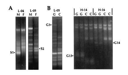
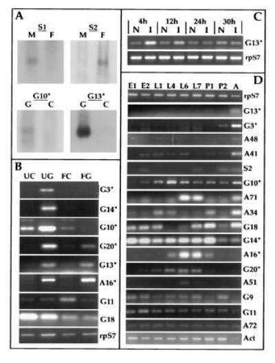
The isolation and study of Anopheles gambiae genes that are differentially expressed in development, notably in tissues associated with the maturation and transmission of the malaria parasite, is important for the elucidation of basic molecular mechanisms underlying vector-parasite interactions. We have used the differential display technique to screen for mRNAs specifically expressed in adult males, females, and midgut tissues of blood-fed and unfed females. We also screened for mRNAs specifically induced upon bacterial infection of larval stage mosquitoes. We have characterized 19 distinct cDNAs, most of which show developmentally regulated expression specificity during the mosquito life cycle. The most interesting are six new sequences that are midgut-specific in the adult, three of which are also modulated by blood-feeding. The gut-specific sequences encode a maltase, a V-ATPase subunit, a GTP binding protein, two different lectins, and a nontrypsin serine protease. The latter sequence is also induced in larvae subjected to bacterial challenge. With the exception of a mitochondrial DNA fragment, the other 18 sequences constitute expressed genomic sequence tags, 4 of which have been mapped cytogenetically.


Figures

Figure 1
Figure 1
Examples of differential display comparing male versus female (A, M versus F) and gut versus carcass (B, G versus C) mRNA. Amplification products were resolved on 1.4% ethidium-stained agarose gels. PCR amplification used decamer primers L-04 (5′-GACTGCACAC-3′), L-05 (5′-ACGCAGGCAC-3′), H-14 (5′-ACCAGGTTGG-3′), and H-16 (5′-TCTCAGCTGG-3′), in combination with an oligo(dT) primer. H-14 and H-16 reactions were performed in duplicate. The differentially amplified PCR fragments S1, S2, G3, G13, and G14 were isolated for further analysis.
Figure 2
Figure 2
mRNA expression analysis of differentially amplified fragments. (A) Northern analysis of adult sex and gut-specific differential display fragments. The female-specific clone S2 as well as amplicon S1 were hybridized to filters containing RNA isolated from unfed females (F) and males (M). The gut-specific clones were hybridized to filters containing RNA from fed (24 h) midguts (G) and remaining carcasses (C). (B) RT–PCR analysis of gut specific and blood meal modulated expression, comparing unfed midguts (UG), carcasses (UC), blood-fed midguts (FG) and carcasses (FC) 22 h postfeeding. cDNA clones are identified on the right margin with asterisks, indicating those shown in Fig. 3. Levels of ribosomal protein S7 RNA were used as control. (C) RT–PCR analysis of G13 mRNA levels in bacterially challenged larvae versus naive larvae at 4, 12, 24, and 30 h following infection. (D) RT–PCR expression analysis comparing RNA from embryos (E1 = 0–24 h; E2 = 28–46 h), larvae (L1 = 0–25 h, L4 = 4 days, L6 = 6 days, L7 = 7 days), pupae (P1 = early and P2 = late), and adult females (A). Ribosomal protein S7 (15) and cytoskeletal actin (16) mRNA levels were used as controls.
Figure 3
Figure 3
Comparisons of deduced amino acid sequences of cloned cDNAs with previously characterized sequences from other organisms. Amino acid identities are indicated by bold type and asterisks. (A) An asterisk indicates the actual C terminus of the protein. First and/or last amino acid positions of the partial sequences are numbered in parentheses. The G3 sequence corresponding to the C-terminal part of a maltase-like protein is aligned with the An. gambiae proteins (17) corresponding to the maltase-like genes (line a) and 1 (line b). The conserved putative catalytic site (NHD) is boxed. (B) The full-length sequence of the putative C-type lectin A16 is aligned with the partial carbohydrate recognition domains of human l-selectin (line a) (18), human macrophage mannose receptor (line b) (19), and Sarcophaga lectin (line c) (20). Motifs characteristic of C-type lectins are boxed (ref. ; A. Ezekowitz and N. Harris, personal communication). (C) The full-length sequence of the putative galactose lectin G20 is aligned with the rat intestinal lactose binding lectin (line a) (22), pig lactose binding lectin (line b) (23), and human Galectin-7 (line c) (24). Conserved amino acids involved in galactose binding are boxed. (D) The full-length sequence of the G10 putative 14K vacuolar ATPase is aligned with the homologous proteins of Drosophila melanogaster (line a) (accession no. S38436S38436), Manduca sexta (line b) (25); and human (line c) (accession no. JC4193). (E) The C-terminal part of the G14 putative low molecular weight G25K GTP binding protein is aligned with the homologous proteins of D. melanogaster (line a) (26) and human (line b) (27). (F) The full-length sequence of the G13 putative serine protease is aligned with Aedes aegypti chymotrypsin (line a) (accession no. U56423U56423), An. gambiae trypsin 7 (line b) (3), mouse mast cell protease 7 (line c) (28), and mouse tissue kallikrein (line d) (29). The residues (H, D, S) of the catalytic triad of serine proteases are underlined. The putative activation peptide (3) is marked with arrows, and the trypsin-specific residues (KD, boxed) (3) of An. gambiae trypsin 7 (sequence b) are boxed.

References

    1. Warburg A, Miller L H. Parasitol Today. 1991;7:179–181. - PubMed
    1. Collins F H, Sakai R K, Vernick K D, Paskewitz S, Seeley D C, Miller L H, Collins W E, Campbell C C, Gwadz R W. Science. 1986;234:607–610. - PubMed
    1. Müller H-M, Catteruccia F, Vizioli J, della Torre A, Crisanti A. Exp Parasitol. 1996;81:371–385. - PubMed
    1. Collins F H. Parasitol Today. 1994;10:370–376. - PubMed
    1. Williams J G K, Hanafey M K, Rafalski J A, Tingey S V. Methods Enzymol. 1993;218:704–740. - PubMed

Publication types

LinkOut - more resources