. 1996 Nov 12;93(23):13164-9.
doi: 10.1073/pnas.93.23.13164.

Induction of apoptosis in rhabdomyosarcoma cells through down-regulation of PAX proteins

Affiliations

Induction of apoptosis in rhabdomyosarcoma cells through down-regulation of PAX proteins

M Bernasconi et al. Proc Natl Acad Sci U S A. .

Abstract

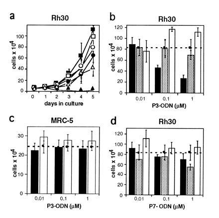
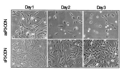
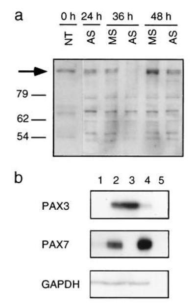
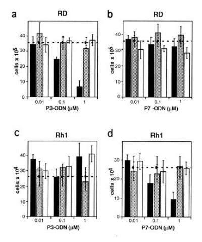
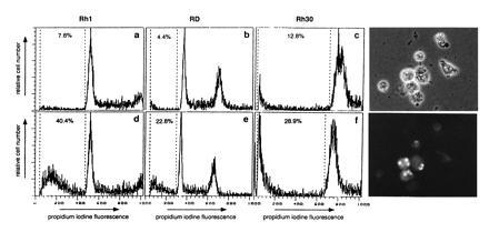
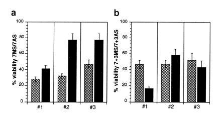
The expression of a number of human paired box-containing (PAX) genes has been correlated with various types of tumors. Novel fusion genes encoding chimeric fusion proteins have been found in the pediatric malignant tumor alveolar rhabdomyosarcoma (RMS). They are generated by two chromosomal translocations t(2;13) and t(1;13) juxtaposing PAX3 or PAX7, respectively, with a forkhead domain gene FKHR. Here we describe that specific down-regulation of the t(2;13) translocation product in alveolar RMS cells by antisense oligonucleotides results in reduced cellular viability. Cells of embryonal RMS, the other major histiotype of this tumor, were found to express either wild type PAX3 or PAX7 at elevated levels when compared with primary human myoblasts. Treatment of corresponding embryonal RMS cells with antisense olignucleotides directed against the mRNA translational start site of either one of these two transcription factors similarly triggers cell death, which is most likely due to induction of apoptosis. Retroviral mediated ectopic expression of mouse Pax3 in a PAX7 expressing embryonal RMS cell line could partially rescue antisense induced apoptosis. These data suggest that the PAX3/FKHR fusion gene and wild-type PAX genes play a causative role in the formation of RMS and presumably other tumor types, possibly by suppressing the apoptotic program that would normally eliminate these cells.


Figures

Figure 1
Figure 1
Cell growth and survival rate in the alveolar RMS cell line Rh30 upon treatment with specific PAX3 antisense oligonucleotides. Rh30 cells express the aRMS specific translocation product PAX3/FKHR. ODN are directed against the translational start site of the PAX3 mRNA. (a) Growth rate of Rh30 cells incubated with different concentrations of antisense PAX3 (as-P3, solid symbols) and sense PAX3 (s-P3, open symbols) ODN. Concentrations used were 0.01 μM (▪), 0.1 μM (•), and 1 μM (▴). Viable cells were counted every 24 hr for five consecutive days. (bd) Viability of Rh30 and control cells incubated with increasing concentrations of antisense (solid bars), missense (shaded bars), or sense (open bars) ODN. The number of cells treated with lipofectin only is indicated by the horizontal lines (dashed lines). Rh30 cells were treated with PAX3 ODN (b) and PAX7 ODN (d), control MRC-5 human lung fibroblasts were treated with PAX3 ODN (c). Cells were counted after 5 days except for in d where they were counted after 3 days. Results shown represent means of at least three independent experiments each using three dishes for every time point counted. Bars = SD.
Figure 2
Figure 2
Representative morphology of Rh30 cells treated with either antisense (as-P3) or sense (s-P3) PAX3 ODN after days 1, 2, and 3. Magnification of the phase contrast picture is ×100.
Figure 3
Figure 3
Expression of PAX protein and mRNA. (a) Western blot analysis of Rh30 cell extracts upon treatment with 1 μM antisense (AS) and missense (MS) PAX3 ODN. An equal number of attached cells at day 0, 1, 1.5, and 2 after ODN incubations were loaded on the gel. The arrow indicates the position of the PAX3/FKHR fusion protein. (b) RNase protection assay was performed using 15 μg total RNA isolated from primary human myoblast cells (lane 1), RD (lane 2), Rh30 (lane 3), Rh1 (lane 4), and yeast (lane 5). Details of the probes used for protection analysis have been described previously (13). The use of a probe from the 5′ end does not allow to distinguish between transcripts derived from the wild-type PAX3 or the translocated PAX3/FKHR gene, however the presence of PAX3/FKHR fusion transcripts and protein in Rh30 cells has been described elsewhere (9, 11). Glyceraldehyde-phosphate-dehydrogenase was used as a control for RNA quantities.
Figure 4
Figure 4
Viability in embryonal RMS cell lines RD (a and b) and Rh1 (c and d) treated with antisense (solid bars), missense (shaded bars), or sense (open bars) oligonucleotides. Viable, attached cells treated with PAX3 (P3, a and c) or PAX7 (P7, b and d) ODN were counted after five days (RD cells) or 3 days (Rh1 cells). The number of cells treated with lipofectin alone is represented by the dashed horizontal line. At least three independent experiments were carried out for each treatment. Bars = SD of the proportion.
Figure 5
Figure 5
Detection of apoptosis after antisense ODN treatment. (af) fluorescence-activated cell sorter cell cycle analysis of the indicated cell lines was performed 60 hr after treatment with 1 μM of either missense ODN (ac Upper) or antisense ODN (df Lower). Attached and floating cells were collected and stained with propidium iodine to reveal DNA content. The percentage of cells below the G1 peak is indicated. (Right) TUNEL labeling of Rh1 cells 24 hr after incubation with 1 μM antisense PAX7 ODN. Phase contrast reveals cells displaying morphological features of apoptosis (Upper), whereas fluorescence microscopy identifies labeled cells (Lower). Quantification of the TUNEL reaction in attached cells reveals that 16.9% (n = 112) of antisense-treated Rh1 cells are labeled vs. 2.5% (n = 159) after sense PAX7 ODN treatment.
Figure 6
Figure 6
Partial suppression of cell death in Rh1 cells upon retroviral mediated ectopic expression of Pax3. After drug selection, pools of Pax3 (solid bars) and control vector (expressing β-galactosidase; shaded bars) transduced cells were incubated with 1 μM of PAX7 antisense (AS) or missense (MS) ODN and viable cells counted after 48 hr (a). (b) The same cells were treated with both PAX3 and PAX7 antisense or missense ODN at 1 μM each. Viable cells were again counted after 48 hr. For both experiments, results of three independent incubations (–3) using at least three dishes each for cell number determination are shown expressed as ratio between MS and AS ODN incubations.

References

    1. Stuart E T, Gruss P. Hum Mol Genet. 1995;4:1717–1720. - PubMed
    1. Strachan T, Read A P. Curr Opin Genet Dev. 1994;4:427–438. - PubMed
    1. Torres M, Gomezpardo E, Dressler G R, Gruss P. Development (Cambridge, UK) 1995;121:4057–4065. - PubMed
    1. Urbanek P, Wang Z, Fetka I, Wagner E F, Busslinger M. Cell. 1994;79:901–912. - PubMed
    1. Hill R, Van Heiningen V. Trends Genet. 1992;8:119–120. - PubMed

Publication types

MeSH terms