. 1996 Nov 12;93(23):13280-5.
doi: 10.1073/pnas.93.23.13280.

Functional breakdown of the lipid bilayer of the myelin membrane in central and peripheral nervous system by disrupted galactocerebroside synthesis

Affiliations

Functional breakdown of the lipid bilayer of the myelin membrane in central and peripheral nervous system by disrupted galactocerebroside synthesis

A Bosio et al. Proc Natl Acad Sci U S A. .

Abstract

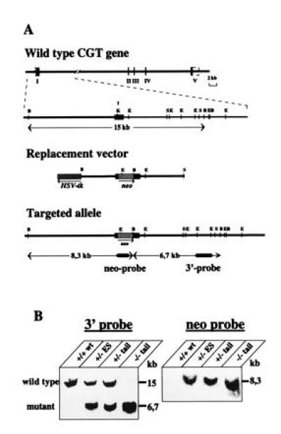
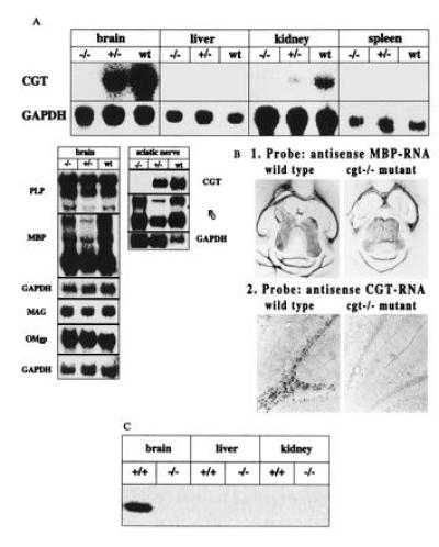
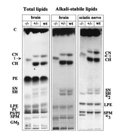
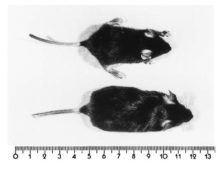
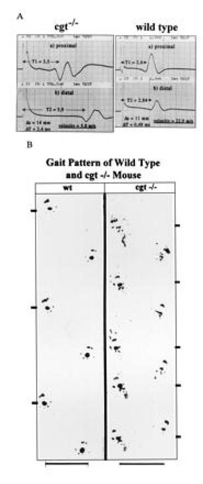
The lipid bilayer of the myelin membrane of the central nervous system (CNS) and the peripheral nervous system (PNS) contains the oligodendrocyte- and Schwann cell-specific glycosphingolipids galactocerebrosides (GalC) and GalC-derived sulfatides (sGalC). We have generated a UDP-galactose ceramide galactosyltransferase (CGT) null mutant mouse (cgt-/-) with CNS and PNS myelin completely depleted of GalC and derived sGalC. Oligodendrocytes and Schwann cells are unable to restore the structure and function of these galactosphingolipids to maintain the insulator function of the membrane bilayer. The velocity of nerve conduction of homozygous cgt-/- mice is reduced to that of unmyelinated axons. This indicates a severely altered ion permeability of the lipid bilayer. GalC and sGalC are essential for the unperturbed lipid bilayer of the myelin membrane of CNS and PNS. The severe dysmyelinosis leads to death of the cgt-/- mouse at the end of the myelination period.


Figures

Figure 1
Figure 1
Disruption of the cgt locus in mouse ES cells and generation of CGT-deficient mice. (A) Targeting strategy: the murine cgt-coding exon I is indicated by a closed box. The neor cassette inserted into the KpnI site of exon I and the HSV-tk gene (▨) has been placed upstream of the exon I. B, BamHI; E, EcoRI; S, SacI; K, KpnI. Homologous recombination introduces an additional BamHI site through the pgk neo cassette. (B) Southern blot hybridization analysis of BamHI-digested ES cells and tail DNA from offsprings of cgt+/− intercrosses. neo probe is a 1.1-kb fragment; 3′-probe a 1.1-kb EcoRI fragment (3) coresponding to a genomic sequence downstream of the replacement sequence.
Figure 2
Figure 2
Analysis of cgt expression and relevant myelin-associated genes of oligodendrocyte by Northern blot, in situ hybridization and enzyme assay. C57BL/6 mice were used as control mice in all comparative experiments. (A) Northern blot analysis of CGT, PLP, MBP, MAG, OMgp, and P0 RNA of brain, liver, kidney, spleen, and sciatic nerve. Approximate sizes of transcripts: CGT, 3.2 kb; PLP, 1.6, 2.4 and 3.2 kb; MBP, 2.1 kb; MAG, 4 kb; OMgp, 1.8 kb; Po, 2.0 kb; glyceraldehyde-phosphate dehydrogenase, 1.2 kb. (B) Comparative in situ hybridization analysis of CGT and MBP expression in wt and homozygous cgt−/− mouse brain. (C) CGT activity of brain, liver and kidney of age matched p18-20 wt and homozygous mutant mice. Enzymatic activity was determined as described (17) with synthetic d-2-hydroxyhexanoylsphingosine and UDP[14C]galactose (Amersham) as substrates and microsomal protein of brain, liver, and kidney from p18 wt and cgt−/− mice. The reaction product, radioactive [14C]galactosyl ceramide, was detected in the lipid extract after separation by TLC as described and recorded with the phosphoimager. Only brain microsomes of wt mouse brain showed significant enzymatic activity, that of kidney was to weak to be detected by autoradiography.
Figure 3
Figure 3
Analysis of myelin lipids of brain and sciatic nerve of wt, hetero-, and homozygous cgt−/− mice (24-day-old). Brain, total and alkali-stable lipids; sciatic nerve, alkali-stable lipids; C, cholesterol; CN, normal fatty acid substituted GalC; CH, α-hydroxy fatty acid substituted GalC; PE, phosphatidylethanolamine; SN, normal fatty acid substituted sGalC; SH, α-hydroxy fatty acid substituted sGalC; LPE, lysophosphatidylethanolamine (lysoplasmalogens); PC, phosphatidylcholine; GM3, ganglioside GM3. Arrows: 1, glucocerebroside; 2, sulfated glucocerebroside; and 3, sphingomyelin-containing long chain α-hydroxy fatty acids.
Figure 4
Figure 4
Age-matched (p24) wt (Lower) and cgt−/− (Upper) mice showing the growth retardation of cgt−/− upper mouse. (Scale in cm.)
Figure 5
Figure 5
(A) Nerve conduction velocity of cgt−/− is decreased from 22–25 m/s to 4–8 m/s when p18-20 mice were compared. (B) Gait pattern of wt and cgt−/− mouse.
Figure 6
Figure 6
Electron microscopy of cross sections of the optic and sciatic nerve of p24 wt, hetero- and homozygous mutant mice. (AC) Optic nerve. Cross section of optic nerve of A, cgt−/−; B, cgt+/−; C, wt. (DF) Sciatic nerve. Cross section of sciatic nerve of D, cgt−/−; E, cgt+/−; F, wt. (Bar = 1.0 mm.)

References

    1. Schulte S, Stoffel W. Proc Natl Acad Sci USA. 1993;90:10265–10269. - PMC - PubMed
    1. Schulte S, Stoffel W. Eur J Biochem. 1995;233:947–953. - PubMed
    1. Bosio A, Binczek E, Stoffel W. Genomics. 1996;35:223–226. - PubMed
    1. Bosio A, Binczek E, Le Beau M M, Fernald A A, Stoffel W. Genomics. 1996;34:69–75. - PubMed
    1. Gard A L, Pfeiffer S E. Neuron. 1990;5:615–625. - PubMed

Publication types

MeSH terms

LinkOut - more resources