. 1996 Dec 1;16(23):7407-15.
doi: 10.1523/JNEUROSCI.16-23-07407.1996.

Cloning and characterization of postsynaptic density 93, a nitric oxide synthase interacting protein

Affiliations

Cloning and characterization of postsynaptic density 93, a nitric oxide synthase interacting protein

J E Brenman et al. J Neurosci. .

Abstract

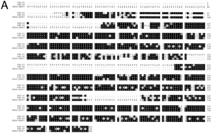
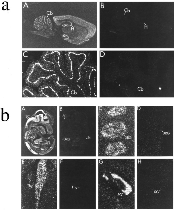
Nitric oxide (NO) formation in brain is regulated by the calcium/calmodulin dependence of neuronal NO synthase (nNOS). Calcium influx through NMDA-type glutamate receptors is efficiently coupled to nNOS activity, whereas many other intracellular calcium pathways are poorly coupled. To elucidate possible mechanisms responsible for this coupling, we performed yeast two-hybrid screening to identify proteins that interact with nNOS. Two nNOS interacting proteins were identified: the postsynaptic density proteins PSD-93 and PSD-95. Here, we report the cloning and characterization of PSD-93. PSD-93 is expressed in discrete neuronal populations as well as in specific non-neuronal cells, and it exhibits complex molecular diversity attributable to tissue-specific alternative splicing. PSD-93, like PSD-95, binds to nNOS and to the NMDA receptor 2B. PSD-93, however, is unique among PSD-95/SAP-90 family members in its expression in Purkinje neuron cell bodies and dendrites. We also demonstrate that the PDZ domain at the N terminus of nNOS is required, but it is not sufficient for interaction with PSD-93/95. Given that PSD-93 and PSD-95 each contain multiple potential binding sites for nNOS and the NMDA receptor, complexes involving oligomers of PSD-93/95 may help account for the functional as well as the physical coupling of nNOS to NMDA receptors.


Figures

Fig. 1.
Fig. 1.
Predicted amino acid sequence and alternative splicing of PSD-93. A, Alignment of PSD-93 with PSD-95 and HDLG-SAP97 indicates an overall amino acid identity of ∼70%. Identical amino acids are indicated by black boxes, and conserved amino acid changes are shaded by gray boxes.B, The N terminus of PSD-93 is alternatively splice prone. Sequences of the four alternative N-terminal transcripts (5′a–d) are indicated. Black triangles denote the sites at which the sequences diverge from PSD-93. The amino acidnumbers in bold refer to that predicted using 5′b of PSD-93. In-frame starter methionines or stop codons are indicated. C, Two different alternatively spliced inserts occur between amino acids K624 and R625. Thearrow indicates the location at which the two isoform insertions occur. The sequences of the alternative insertions are also shown. D, A schematic model showing the domain structure of PSD-93. Identified sites of putative alternative splicing are indicated. (PSD-93 has been given GenBank accession number U50717.)Figure continues.
Fig. 1.
Fig. 1.
Predicted amino acid sequence and alternative splicing of PSD-93. A, Alignment of PSD-93 with PSD-95 and HDLG-SAP97 indicates an overall amino acid identity of ∼70%. Identical amino acids are indicated by black boxes, and conserved amino acid changes are shaded by gray boxes.B, The N terminus of PSD-93 is alternatively splice prone. Sequences of the four alternative N-terminal transcripts (5′a–d) are indicated. Black triangles denote the sites at which the sequences diverge from PSD-93. The amino acidnumbers in bold refer to that predicted using 5′b of PSD-93. In-frame starter methionines or stop codons are indicated. C, Two different alternatively spliced inserts occur between amino acids K624 and R625. Thearrow indicates the location at which the two isoform insertions occur. The sequences of the alternative insertions are also shown. D, A schematic model showing the domain structure of PSD-93. Identified sites of putative alternative splicing are indicated. (PSD-93 has been given GenBank accession number U50717.)Figure continues.
Fig. 2.
Fig. 2.
Tissue expression and alternative splicing of PSD-93. A, Northern blotting of adult rat tissues indicates that PSD-93 is expressed as an ∼7.5 kb transcript that occurs in brain (Br) but not in kidney (Ki), spleen (Sp), liver (Li), heart (He), or skeletal muscle (Sk). C, Northern blotting of brain regions demonstrates that PSD-93 is present in cerebellum (Cb), cortex (Cx), hippocampus (Hi), and striatum (St) but is absent from brainstem (BS). Note that the band in cerebellum migrates slightly faster than that in other brain regions. ForA and B, a probe common to all of the alternatively spliced forms of PSD-93 was used. D, E, The blot in B was sequentially rehybridized with probes corresponding to two of the alternatively spliced N-terminal regions of PSD-93. C, Probing with 5′a reveals that the regional distribution of this splice variant is similar to that of PSD-93.D, 5′b, however, is selectively absent from cerebellum.B, F, Duplicate samples of mRNA were probed for glyceraldehyde 3-phosphate dehydrogenase to demonstrate loading and integrity of the mRNA for the above blots.
Fig. 3.
Fig. 3.
Cellular localization of PSD-93 mRNA in adult brain and E15 embryo. In situ hybridization was used to localize transcripts for PSD-93 (A, C, E, G) or sense control (B, D, F, H). a, In adult brain, PSD-93 seems to be neuron-specific and is highly expressed in Purkinje neurons in the cerebellum (Cb) and also occurs in pyramidal and granule cells in hippocampus (H). b, In E15 embryo, PSD-93 is abundantly expressed in neurons of spinal cord (SC), dorsal root ganglia (DRG), and intestine (In). PSD-93 is also observed in cells of the thymus (Thy) and submandibular gland (SG).
Fig. 4.
Fig. 4.
Specificity of antisera to PSD-93 and PSD-95. Western blot analysis reveals that the predominant PSD-93 protein product in rat brain migrates at 103 kDa, whereas the major PSD-95 reactive band migrates at 95 kDa. Crude adult rat brain homogenates (50 μg of protein/lane) were size-fractionated by SDS-PAGE and analyzed by immunoblotting with affinity-purified antiserum to either PSD-93 (lane 1) or PSD-95 (lane 2). Size markers are in kilodaltons.
Fig. 5.
Fig. 5.
PSD-93 is postsynaptic, whereas PSD-95 is predominantly presynaptic in rat cerebellum. A sagittal section of rat cerebellum was processed for indirect immunofluorescent double-labeling using a guinea pig antiserum to PSD-93 and a rabbit antiserum to PSD-95. A, PSD-93 immunoreactivity, visualized in thered channel, is present in Purkinje cell somata and molecular layer (M) of cerebellum (100× magnification). B, PSD-95, visualized in thegreen channel, is present in the synaptic plexus of basket cell axons beneath the Purkinje cell layer (P) (100× magnification). C, Higher-power double exposure shows that PSD-93 immunoreactivity (orange) is confined to Purkinje neurons (arrow), whereas PSD-95 immunoreactivity (green) primarily labels the presynaptic basket cell pinceaus (arrowhead) (400× magnification). Note that the orange colorobserved on double exposure is attributable to the longer exposure times required by FITC filters.
Fig. 6.
Fig. 6.
The PDZ repeats of PSD-93 interact with nNOS and the tSXV motif of NMDA receptor 2B. Glutathione-Sepharose beads bound to GST or to a PSD-93 protein fragment (amino acids 77–453) fused toGST (G-P93) were incubated with brain extracts. After the beads were washed extensively, retained proteins were eluted with 0.2% SDS and separated by SDS/PAGE. Western blotting indicates that nNOS is selectively retained by G-P93 (lane 3) but not by GST alone (lane 2). Binding assays performed in parallel indicate that an NMDA receptor 2B C-terminal peptide displaces nNOS from G-P93 completely at 10 μm(lanes 4 and 5), substantially at 5 μm (lanes 6 and 7), and negligibly at 0.1 μm (lanes 8 and9). A control peptide had no effect on nNOS binding to G-P93 at 10 μm (lanes 10 and11). Input = 10% of the protein loaded onto the fusion-protein columns.
Fig. 7.
Fig. 7.
The 100 amino acid PDZ domain of nNOS is not sufficient to bind to PSD-93. Glutathione-Sepharose beads bound to GST alone or to GST fusion-protein fragments encoding various N-terminal domains of nNOS (G-nNOS) were incubated with brain extracts. After the beads were washed extensively, retained proteins were eluted with 0.2% SDS and separated by SDS/PAGE. Western blotting with PSD-93 antiserum reveals that PSD-93 does not bind to eitherGST alone (lane 2) or toG-nNOS1-100 (lane 3) but is selectively retained by G-nNOS1-150 (lane 4) and G-nNOS1-195 (lane 5). Input = 10% of the protein loaded onto the fusion-protein columns (lane 1).

References

    1. Aoki C, Fenstemaker S, Lubin M, Go CG. Nitric oxide synthase in the visual cortex of monocular monkeys as revealed by light and electron microscopic immunocytochemistry. Brain Res. 1993;620:97–113. - PubMed
    1. Bredt DS, Snyder SH. Nitric oxide mediates glutamate-linked enhancement of cGMP levels in the cerebellum. Proc Natl Acad Sci USA. 1989;86:9030–9033. - PMC - PubMed
    1. Bredt DS, Snyder SH. Isolation of nitric oxide synthetase, a calmodulin-requiring enzyme. Proc Natl Acad Sci USA. 1990;87:682–685. - PMC - PubMed
    1. Bredt DS, Snyder SH. Nitric oxide, a novel neuronal messenger. Neuron. 1992;8:3–11. - PubMed
    1. Brenman JE, Chao DS, Xia H, Aldape K, Bredt DS. Nitric oxide synthase complexed with dystrophin and absent from skeletal muscle sarcolemma in Duchenne muscular dystrophy. Cell. 1995;82:743–752. - PubMed

Publication types

MeSH terms

Associated data

LinkOut - more resources