. 1996 Dec 1;16(23):7437-46.
doi: 10.1523/JNEUROSCI.16-23-07437.1996.

Ultrastructural localization of dendritic messenger RNA in adult rat hippocampus

Affiliations

Ultrastructural localization of dendritic messenger RNA in adult rat hippocampus

M E Martone et al. J Neurosci. .

Abstract

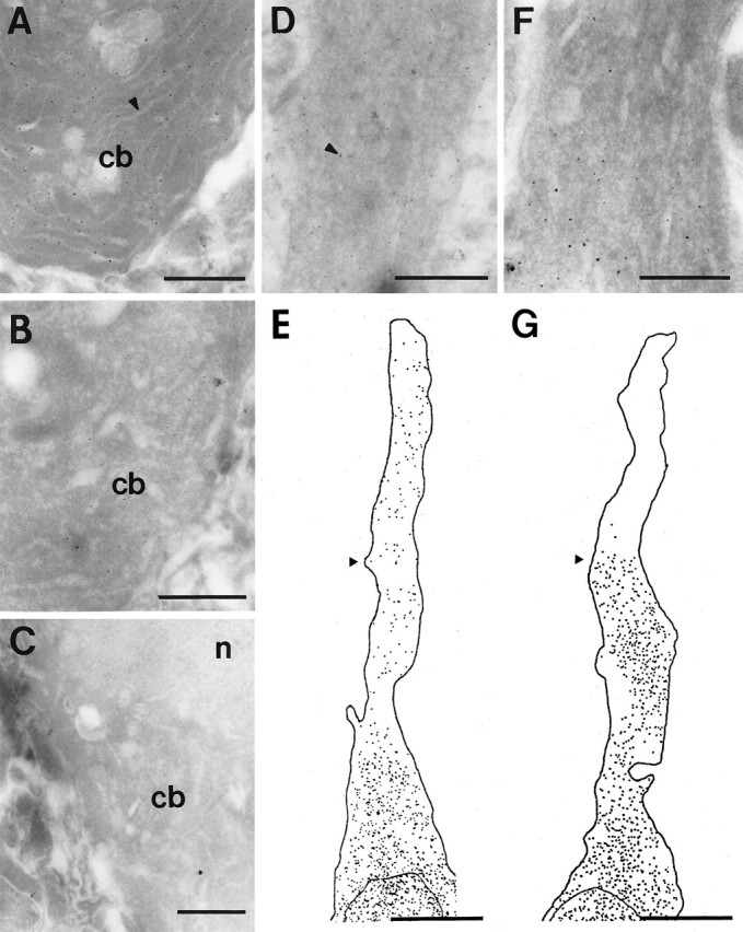
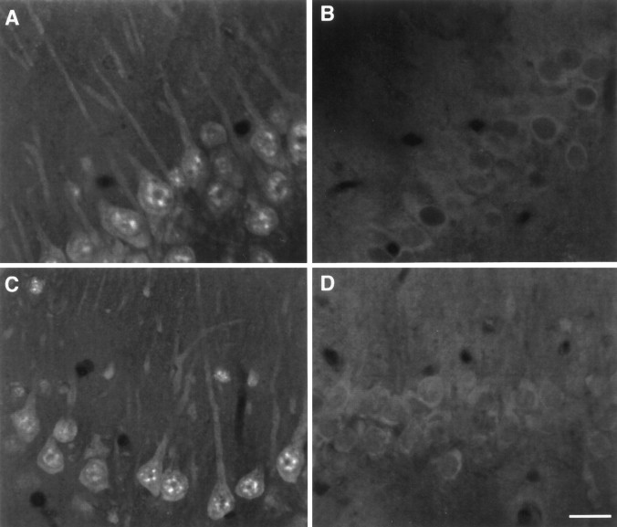
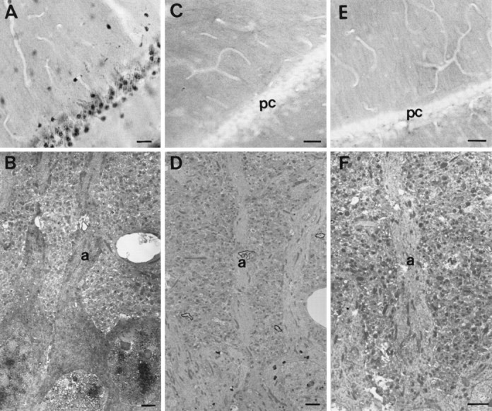
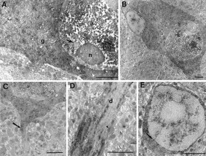
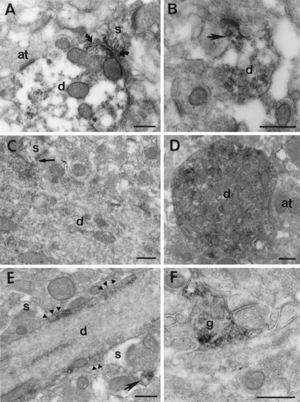
An ultrastructural examination of mRNA within adult rat CA1 hippocampal dendrites was conducted using two different methods. The messages for the alpha and beta forms of the calcium-calmodulin-dependent protein kinase II were localized in ultracryosections using silver-intensified gold detection of isoform-specific oligonucleotide probes. Labeling for both isoforms was observed within the cell bodies and proximal dendrites of pyramidal neurons, but only the alpha form was observed in more distal dendrites. Unfortunately, the morphological preservation of the tissue was not sufficient to determine the localization of labeling relative to subcellular features such as dendritic spines. To address this issue, a preembedding peroxidase-based method was developed, resulting in better preservation of the neuropil. The total population of polyadenylated [poly(A)] mRNA was localized in hippocampus using a biotinylated poly(dT) probe. Poly(A) mRNA was present in the nucleus and throughout the cell body of all hippocampal cells and within isolated dendrites and glial processes within the neuropil. Within pyramidal neurons, labeling was distributed in a longitudinal pattern in proximal apical dendrites. More distally, the amount of labeling diminished, and smaller foci of labeling were observed, particularly near the plasma membrane. Concentrated labeling was present at the base of dendritic spines and, less frequently, near synapses onto the dendritic shaft. These results suggest that dendritic mRNA is found in the vicinity of postsynaptic sites and provide additional evidence that local protein synthesis may play an important role in establishing and maintaining synaptic specializations.


Figures

Fig. 1.
Fig. 1.
Localization of mRNA for CAMKII in 1-μm-thick ultracryosections of CA1 pyramidal neurons hybridized with either the 600 bp cDNA probe (A) or the α- or β-specific probes (DG). A, Labeling with the 600 bp CAMKII cDNA probe in a pyramidal cell body (cb). Subcellular features such as the endoplasmic reticulum (arrowhead) were visible in such sections, although membrane contrast was poor. B, Control section hybridized with a 500 bp fragment of bacteriophage λ. A small amount of labeling is present within the cell body (cb).C, Control section in which the probe was omitted from the hybridization solution. Little gold labeling was observed in the cell body (cb) or nucleus (n).D, E, Labeling for the α CAMKII mRNA in the cell body and apical dendrite of a pyramidal neuron. A tracing of the distribution of silver particles in a montage composed of four micrographs is shown in E. A micrograph through the portion of the dendrite indicated by the arrowhead is shown in D; the arrowhead points to a silver-intensified gold particle. Note that that labeling is fairly evenly distributed throughout the apical dendrite. F,G, The distribution of β CAMKII mRNA in the apical dendrite of a pyramidal neuron. A tracing of the distribution of silver particles in a montage through the apical dendrite is shown inG. Note that the labeling abruptly stops at a distance of ∼15 μm from the cell body. A micrograph through the region indicated by the arrowhead is shown in F. Scale bars: AD, F, 1 μm; E, G, 10 μm.
Fig. 2.
Fig. 2.
Fluorescence in situ hybridization of poly(A) mRNA in pyramidal neurons of area CA1. A,C, Sections labeled for poly(A) mRNA. Labeling could be followed within apical dendrites for a distance of >100 μm.B, Control section hybridized with a poly(dA) sense probe. D, Control section in which no probe was included in the hybridization cocktail. Scale bar, 20 μm.
Fig. 3.
Fig. 3.
Peroxidase labeling for poly(A) mRNA labeling in area CA1 in experimental and control conditions at the light microscopic (A, C, E) and electron microscopic (B, D,F) levels. A, B, Pyramidal neurons labeled for poly(A) mRNA. At both the LM and EM levels, staining was observed within cell bodies, nuclei, and apical dendrites (a). C, D, Pyramidal cell layer (pc) hybridized with the sense probe. E, F, No probe control. In neither control condition was labeling observed within the pyramidal cell layer (pc) or apical dendrites (a). All electron micrographs were taken from the surface of the tissue. Scale bars: A, C,E, 100 μm; B, D,F, 2 μm.
Fig. 4.
Fig. 4.
Ultrastructural localization of poly(A) mRNA in cell bodies and dendrites of rat hippocampal cells. A, Labeled pyramidal cell body. Both densely labeled foci and a more diffuse background staining were observed in the nucleus, although neither the nucleolus (n) nor the heterochromatin (arrow) was labeled. Labeling was present in a finer punctate pattern in the cytoplasm that was not associated with the Golgi apparatus (g) or mitochondria.B, Large neuron flanked on the left by a smaller satellite cell. The nuclear speckles were larger and more intensely labeled in the large neurons compared with smaller glial cells. C, Axon initial segment of a labeled pyramidal neuron (arrow). Labeling extended into the beginning portion but then terminated. D, Pyramidal cell dendrite (d). Labeling was distributed in a roughly longitudinal pattern within heavily labeled dendrites. Thearrowhead points to a concentration of labeling underneath a synaptic apposition onto the dendritic shaft.E, Small, likely glial, cell showing the distribution of poly(A) mRNA within the nucleus. In this cell, tracks of mRNA radiate from the interior of the nucleus to the nuclear membrane and perhaps into the cytoplasm (arrow). Scale bars, 1 μm.
Fig. 5.
Fig. 5.
Ultrastructural localization of poly(A) mRNA in isolated dendrites and processes within the neuropil. A, Cross-section of a spiny dendrite (d) with a large spine (s) attached. Intense labeling is present associated with a membranous profile at the base of the spine (curved arrows). Little labeling is present near the axonal terminal (at) onto the dendritic shaft. B, Cross-section of a small spiny dendrite (d) with a labeled spine (arrow) attached. C, Longitudinal labeled dendrite (d) with a spine (s) attached. Concentrated labeling and a profile of endoplasmic reticulum are present at the base of the spine (arrow). This tissue was counterstained with uranyl acetate and lead, which obscured the labeling to some extent.D, Cross section of a labeled dendrite (d) with an axon terminal (at) forming a symmetrical synapse. The heaviest staining within the dendrite is not associated with the synaptic site. E, Longitudinal labeled dendrite (d) with two spines (s) nearby but not attached. Labeling within the dendrite is associated with slight bulges in the plasma membrane (arrowheads), which may represent sites of attachment of dendritic spines that are out of the plane of section. The spine to the lower right shows labeling (arrow) in what could be a spine neck attached to a nearby dendrite. F, Labeling of a glial process (g) apposed to a synaptic complex in the neuropil. Scale bars, 500 nm.

References

    1. Ainger K, Avossa D, Morgan F, Hill SJ, Barry C, Barbarese E, Carson JH. Transport and localization of exogenous myelin basic protein mRNA microinjected into oligodendrocytes. J Cell Biol. 1993;123:431–441. - PMC - PubMed
    1. Bassell GJ, Singer RH, Kosik KS. Association of poly(A) mRNA with microtubules in cultured neurons. Neuron. 1994;12:571–582. - PubMed
    1. Burgin KE, Waxham MN, Rickling S, Westgate SA, Mobley WC, Kelly PT. In situ hybridization histochemistry of Ca2+/calmodulin-dependent protein kinase in developing rat brain. J Neurosci. 1990;10:1788–1798. - PMC - PubMed
    1. Chicurel ME, Harris KM. Three-dimensional analysis of the structure and composition of CA3 branched dendritic spines and their relationship with mossy fiber boutons in the rat hippocampus. J Comp Neurol. 1992;325:169–182. - PubMed
    1. Chicurel ME, Terrian DM, Potter H. mRNA at the synapse: analysis of synaptosomal preparation enriched in hippocampal dendritic spines. J Neurosci. 1993;13:4054–4063. - PMC - PubMed

Publication types

Substances

LinkOut - more resources