. 1996 Dec 1;16(23):7458-68.
doi: 10.1523/JNEUROSCI.16-23-07458.1996.

Two alternatively spliced forms of the cGMP-gated channel alpha-subunit from cone photoreceptor are expressed in the chick pineal organ

Affiliations

Two alternatively spliced forms of the cGMP-gated channel alpha-subunit from cone photoreceptor are expressed in the chick pineal organ

W Bönigk et al. J Neurosci. .

Abstract

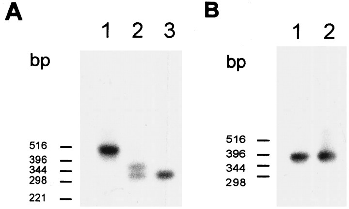
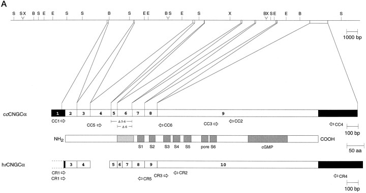
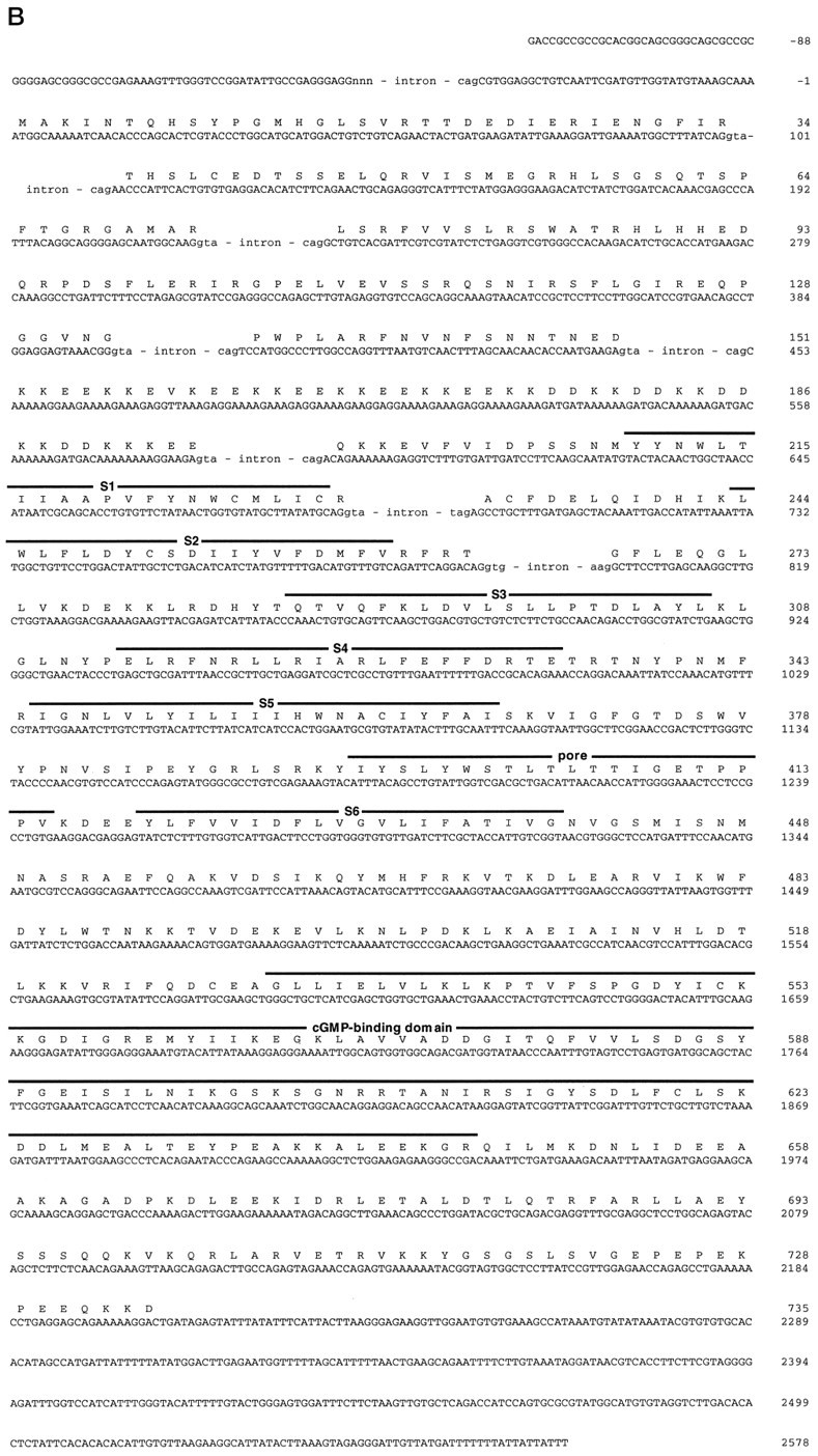
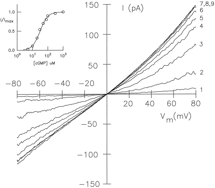
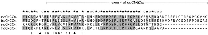
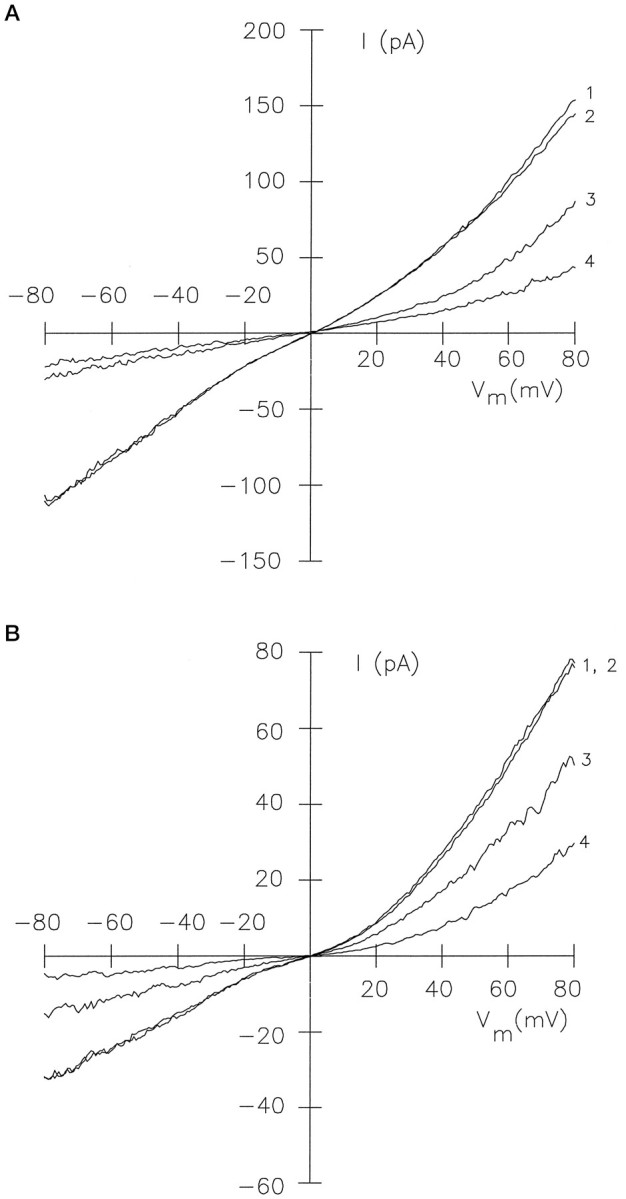
Light sensitivity of the pineal has been retained in most vertebrates, except mammals. Retinal photoreceptors and pinealocytes share common components of light-dependent signaling pathways. In particular, an ion channel gated by cGMP has been electrophysiologically identified in chick pinealocytes; however, the physiological function of a light-sensitive enzyme cascade is not known, and primary structures of only a few pineal components have been determined. By PCR analysis and cloning of the respective cDNA, we show that the chick pineal expresses the alpha-subunit of the cyclic nucleotide-gated (CNG) channel of rod photoreceptors and two short forms of the cone CNG channel. Analysis of the chick cone CNG channel gene reveals that these forms are produced by alternative splicing, which removes either one or two exons from the transcript. The shorter splice variant is functional when heterologously expressed, and it is approximately twofold more sensitive to activation by cGMP than the cone CNG channel. The chick cone CNG channel and the pineal splice form are both modulated by Ca2+/calmodulin (CaM). The CaM sensitivity might be mediated by a putative CaM-binding site in an N-terminal segment encoded by exon 4. This exon is missing in the gene for the rod CNG channel alpha-subunit. Pineal CNG channels are candidates for receptor-mediated Ca2+ entry into pinealocytes and may be an important element of signaling pathways that control the light response and secretion of the pineal hormone melatonin.


Figures

Fig. 2.
Fig. 2.
Transcript structure of ccCNGCα and crCNGCα. Blot hybridization of PCR fragments amplified from cDNA with ccCNGCα-specific (A) and crCNGCα-specific (B) primer pairs (CC5/CC6 and CR1/CR5, respectively). The blot was hybridized with radioactively labeled DNA probes amplified with the same primer set and using cDNA clones pCCG6 and pCCG8B (Bönigk et al., 1993) as templates. Lanes inA: 1, retina; 2, pineal;3, testis. Lanes in B:1, retina; 2, pineal. For negative control PCR, no hybridization signals were detected (data not shown).
Fig. 1.
Fig. 1.
Gene structure of the chick cone CNG channel.A, Top, Restriction map of chromosomal DNA encompassing exons 1–9 of the chick cone CNG channel gene.B, BamHI; E,EcoRI; S, SacI;X, XbaI. A, Bottom, Comparison of the exon structure of the chick cone CNG channel (ccCNGCα) and the human rod CNG channel (hrCNGCα; Dhallan et al., 1992) with the transmembrane topology of CNG channels. Solid andopen boxes, respectively, refer to noncoding and coding regions of exons. Gray boxes refer to transmembrane segments S1–S6, the pore region, and the cGMP-binding site (cGMP); the striped box indicates the highly charged domain in the N-terminal region of the polypeptide. Thearrows refer to primer pairs CC1/CC2, CC3/CC4, and CC5/CC6 used for amplification of cone-specific cDNA fragments, or to the primer pairs CR1/CR2, CR3/CR4, and CR1/CR5 used for amplification of rod-specific cDNA fragments. B, Nucleotide sequence (with splice junction donor and acceptor sequences of exon/intron boundaries in the cDNA) and deduced amino acid sequence of ccCNGCα. Intron sequences at intron/exon boundaries are given in small letters. Numbering of nucleotide positions refers to the cDNA sequence and begins with +1 at the adenine nucleotide of the start codon. Figure continues.
Fig. 1.
Fig. 1.
Gene structure of the chick cone CNG channel.A, Top, Restriction map of chromosomal DNA encompassing exons 1–9 of the chick cone CNG channel gene.B, BamHI; E,EcoRI; S, SacI;X, XbaI. A, Bottom, Comparison of the exon structure of the chick cone CNG channel (ccCNGCα) and the human rod CNG channel (hrCNGCα; Dhallan et al., 1992) with the transmembrane topology of CNG channels. Solid andopen boxes, respectively, refer to noncoding and coding regions of exons. Gray boxes refer to transmembrane segments S1–S6, the pore region, and the cGMP-binding site (cGMP); the striped box indicates the highly charged domain in the N-terminal region of the polypeptide. Thearrows refer to primer pairs CC1/CC2, CC3/CC4, and CC5/CC6 used for amplification of cone-specific cDNA fragments, or to the primer pairs CR1/CR2, CR3/CR4, and CR1/CR5 used for amplification of rod-specific cDNA fragments. B, Nucleotide sequence (with splice junction donor and acceptor sequences of exon/intron boundaries in the cDNA) and deduced amino acid sequence of ccCNGCα. Intron sequences at intron/exon boundaries are given in small letters. Numbering of nucleotide positions refers to the cDNA sequence and begins with +1 at the adenine nucleotide of the start codon. Figure continues.
Fig. 3.
Fig. 3.
Immunohistochemical localization of cone CNG channel in chick and bovine pineal organ. A, Horizontal cryostat section (12 μm) of the chick pineal organ stained with antibody 63–4 and visualized by peroxidase reaction. Cells of two pineal follicles are labeled, whereas pinealocytes outside the follicle are stained less prominently. Scale bar, 20 μm. B, Same as in A at higher magnification. Scale bar, 10 μm. C, Horizontal paraffin section (6 μm) of the bovine pineal organ stained with antibody PPc15 and visualized by peroxidase reaction. PPc15 immunoreactivity is observed in only a few cells in the cortex of the pineal, whereas cells of the medullary part of the organ are not labeled. Scale bar, 20 μm. D, Same as in C at higher magnification. Scale bar, 10 μm. E, Control section of chick pineal. Specific staining is abolished after preincubation of antibody 63–4 with the respective antigenic peptide (20 μg/ml). Scale bar, 20 μm.F, Control section of bovine pineal. No staining was observed when the primary antibody PPc15 was omitted. Picture was taken with Nomarski optics. Scale bar, 20 μm.
Fig. 4.
Fig. 4.
Activation of splice variant ccCNGCαΔ5-6 by cGMP. Series ofIV recordings in the presence of different cGMP concentrations. Inside-out patch of HEK 293 cells transfected with plasmid pccCNGCαΔ5-6. The inset shows the dose–response relation at −60 mV. TheK½ value was 30 μm, and the Hill coefficient was n = 1.8. The cGMP concentrations were trace 1, 5 μm; 2, 10 μm; 3, 20 μm;4, 30 μm; 5, 50 μm; 6, 80 μm;7, 100 μm; 8, 300 μm; 9, 1000 μm.
Fig. 5.
Fig. 5.
Sequence comparison of regions containing a CaM-binding motif in cone and olfactory CNG channels. Sequence alignment of exon 4-encoded region of ccCNGCα with a homologous region of CNG channels from bovine cone photoreceptor (bcCNGCα; Weyand et al., 1994), rat olfactory epithelium (roCNGCα; Dhallan et al., 1990), and fish olfactory epithelium (foCNGCα; Goulding et al., 1992).Arrowheads indicate the two aromatic or long-chain amino acid residues; + indicates the basic residues; and hrefers to hydrophobic residues in the amphiphilic domain of the CaM-binding site. The stars above the sequences indicate that at least three residues at this position are identical;open circles indicate that at least three residues at this position are conserved.
Fig. 6.
Fig. 6.
Modulation of channel activity by Ca2+/CaM. A, Effect of Ca2+/CaM on the cGMP-activated current of ccCNGCα expressed inXenopus oocytes (inside-out patch). The bath solutions contained 120 mm K+-gluconate, 10 mm HEPES-KOH, 50 μm Ca2+, and the following additions: 500 μm cGMP (trace 1); 500 μm cGMP and 1.2 μm CaM (trace 2); 20 μm cGMP (trace 3); 20 μm cGMP and 1.2 μm CaM (trace 4). B, Effect of Ca2+/CaM on the cGMP-activated current of splice variant ccCNGCαΔ5-6 expressed in HEK 293 cells. Same conditions as in A except that the CaM concentration was 600 nm, and the solution was based on KCl instead of K+-gluconate.

References

    1. Abe T, Nakabayashi H, Tamada H, Takagi T, Sakuragi S, Yamaki K, Shinohara T. Analysis of the human, bovine and rat 33 kDa proteins and cDNA in retina and pineal gland. Gene. 1990;91:209–215. - PubMed
    1. Abe T, Shinohara T. S-antigen from the rat retina and pineal gland have identical sequences. Exp Eye Res. 1990;51:111–112. - PubMed
    1. Altenhofen W, Ludwig J, Eismann E, Kraus W, Bönigk W, Kaupp UB. Control of ligand specificity in cyclic nucleotide-gated channels from rod photoreceptors and olfactory epithelium. Proc Natl Acad Sci USA. 1991;88:9868–9872. - PMC - PubMed
    1. Baumann A, Frings S, Godde M, Seifert R, Kaupp UB. Primary structure and functional expression of a Drosophila cyclic nucleotide-gated channel present in eyes and antennae. EMBO J. 1994;13:5040–5050. - PMC - PubMed
    1. Bönigk W, Altenhofen W, Müller F, Dose A, Illing M, Molday RS, Kaupp UB. Rod and cone photoreceptor cells express distinct genes for cGMP-gated channels. Neuron. 1993;10:865–877. - PubMed

Publication types

LinkOut - more resources