. 1996 Dec 1;16(23):7469-77.
doi: 10.1523/JNEUROSCI.16-23-07469.1996.

Intrinsic injury signals enhance growth, survival, and excitability of Aplysia neurons

Affiliations

Intrinsic injury signals enhance growth, survival, and excitability of Aplysia neurons

R T Ambron et al. J Neurosci. .

Abstract

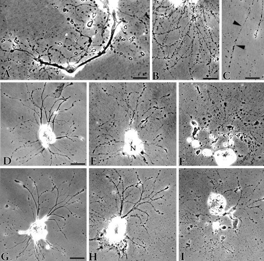
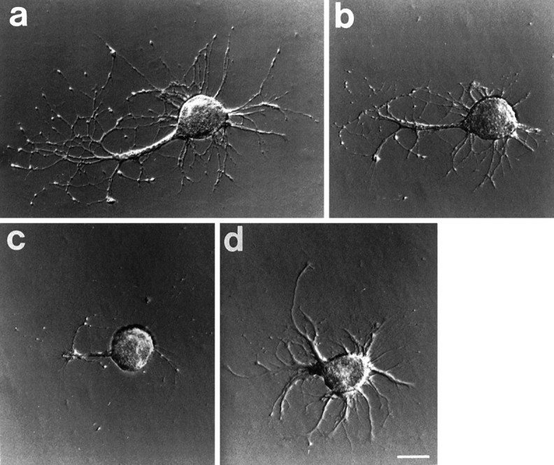
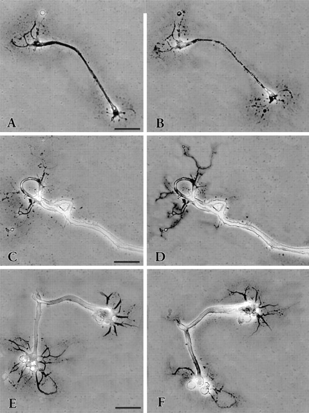
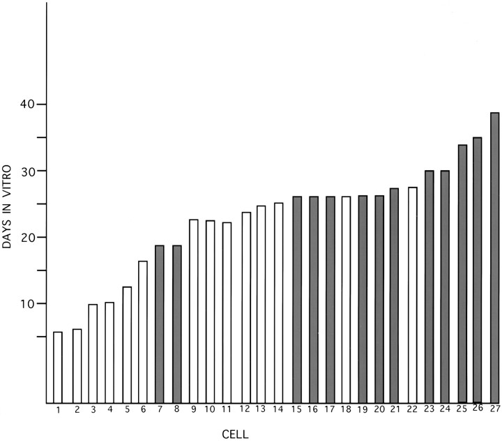
Neurons undergo extensive changes in growth and electrophysiological properties in response to axon injury. Efforts to understand the molecular mechanisms that initiate these changes have focused almost exclusively on the role of extrinsic signals, primarily neurotrophic factors released from target and glial cells. The objective of the present investigation was to determine whether the response to axonal injury also involves intrinsic axoplasmic signals. Aplysia neurons were removed from their ganglia and placed in vitro on a substratum permissive for growth, but in the absence of glia and soluble growth factors. Under these conditions, neurites emerged and grew for approximately 4 d. Once growth had ceased, the neurites were transected. In all, 46 of 50 cells regenerated, either by resorbing the remaining neurites and elaborating a new neuritic arbor or by merely replacing the neurites that had been severed. Cut cells also exhibited enhanced excitability and, paradoxically, prolonged survival, when compared with uninjured neurons. These findings indicate that axons contain intrinsic molecular signals that are directly activated by injury to trigger changes underlying regeneration and compensatory plasticity.


Figures

Fig. 1.
Fig. 1.
Phase-contrast views depicting the fate ofAplysia neurons in vitro.A, Neuritic processes 5 d after plating showing the bulbous, birefringent endings that have replaced the growth cones on the newly formed neurites. The appearance of these structures marks the end of the growth period. Scale bar, 50 μm. B, Cell death typically ensues when the neurites become varicose. Scale bar, 50 μm. C, The varicosities are connected via fine strands (arrowheads). These eventually break, resulting in fragmentation of the neurite. Scale bar, 30 μm. The progression of two cells toward death is shown in DFand GI. The cells were photographed 1 d after plating (D, G) and then 4 d later (E, H) when the neurites were becoming varicose. The nucleus (N) in E has moved to one pole of the cell. After an additional 9 d (F) and 8 d (I), each cell had disintegrated. The entire process from plating to death takes 17 d on average. Scale bar for both cells, 100 μm.
Fig. 2.
Fig. 2.
Phase-contrast photomicrographs of neurons showing the two typical patterns of neurite regeneration. A, An example of regenerative growth in which the new growth merely extends the cut neurites 4 d after plating growth had ceased and the neurites were cut at the arrowhead. Fifteen minutes later the cell was photographed again (C). One day later (F) several prominent growth cones were visible. In the ensuing days the neurites extended, retracted, and then slowly extended again. Seventeen days later, the cell was still growing (I), and it survived >32 d. The other response (B) is much more complex. The neurites of this cell were cut after 4 d in culture (arrowheads), and the cell was photographed 15 min later (D). During the next 11 d the neurite stumps were resorbed into the soma, and new growth emerged (G). The neurites formed thick fascicles that grew extensively and by 24 d after cutting had formed an arbor very different from the original (J) (see also Fig. 3). This cell survived for 31 d. Scale bar, A, B, 100 μm. A control cell (E) in the dish with the cell in A had stopped growing after 4 d. Varicosities on the neurite began to appear 6 d later (H); by day 17 many of the neurites had disappeared, and the cell began to disintegrate (K). Scale bar, 150 μm.
Fig. 3.
Fig. 3.
Photomicrographs of a cell after transecting neurites in vitro. a, The cell is seen 9 d after plating and just before the neurites were cut. Neurites extend from the large remnant of the original axon. b, The cell 15 min after the neurites had been severed and (c) 7 d later. The appearance is radically altered because the axon and many of the neurites were resorbed.d, After an additional 7 d, new neurites have emerged. These continued to grow for several more days, and the cell died on day 35. Scale bar, 100 μm.
Fig. 4.
Fig. 4.
Typical examples of neurite outgrowth from isolated axons. Axons, which had detached from their cell bodies during plating, sprouted neurites within the first 24 hr (A, C, E), but there was little subsequent growth. The axons were followed for 22 d (B), 9 d (D), and 4 d (F), respectively; shortly thereafter, each disintegrated. Scale bar, 100 μm.
Fig. 5.
Fig. 5.
Histogram showing the increased survival time after axotomy in vitro. Twenty-seven cells growing in four dishes in vitro were followed on a daily basis until they died. The neurites of some cells, selected at random, were severed after growth had ceased (filled bars), and their survival was compared with control cells in the same dish (open bars).
Fig. 6.
Fig. 6.
Long-term hyperexcitability induced by transecting neurites of isolated sensory and motor neurons in the presence or absence of hemolymph in the culture medium. A, Examples of repetitive firing during testing of a control sensory neuron and a sensory neuron, the outgrowing neurites of which had been transected 1 d earlier. The test stimulus was a 1 sec depolarizing pulse injected into the neuronal soma through the recording electrode. Injected current was set at 2.5 times that required to reach spike threshold during a previous series of 20 msec pulses. B, Repetitive firing in sensory neurons 1 d after neurite transection. The mean ± SEM number of spikes evoked by the test stimulus was enhanced by previous transection but unaffected by the presence or absence of hemolymph during the test. Hemolymph present,n = 12 control and 10 transected cells,p < 0.005; hemolymph absent, n= 14 control and 6 transected neurons, p < 0.005.C, Repetitive firing in motor neurons 1 d after neurite transection. Firing was enhanced by previous transection and unaffected by hemolymph during the test. Hemolymph present,n = 6 control and 3 transected cells,p < 0.05; hemolymph absent, n= 8 control and 8 transected neurons, p < 0.05.

References

    1. Alberini CM, Ghirardi M, Metz R, Kandel ER. C/EBP is an immediate-early gene required for the consolidation of long-term facilitation in Aplysia. Neuron. 1994;76:1099–1114. - PubMed
    1. Alberini CM, Ghirardi M, Huang Y-Y, Nguyen PV, Kandel ER. A molecular switch for the consolidation of long-term memory: cAMP-inducible gene expression. Ann NY Acad Sci. 1995;256:261–286. - PubMed
    1. Ambron RT, Walters ET. Priming events and retrograde injury signals: a new perspective on the cellular and molecular biology of nerve regeneration. Mol Neurobiol. 1996;13:61–79. - PubMed
    1. Ambron RT, Goldman JE, Schwartz JH. Effect of inhibiting protein synthesis on axonal transport of membrane glycoproteins in an identified neuron of Aplysia. Brain Res. 1975;94:307–323. - PubMed
    1. Ambron RT, Dulin MF, Zhang X-P, Schmied R, Walters ET. Axoplasm enriched in a protein mobilized by nerve injury induces memory-like alterations in Aplysia neurons. J Neurosci. 1995;15:3440–3446. - PMC - PubMed

Publication types

LinkOut - more resources