. 1996 Nov 15;16(22):7117-27.
doi: 10.1523/JNEUROSCI.16-22-07117.1996.

Molecular determinants of beta 1 subunit-induced gating modulation in voltage-dependent Na+ channels

Affiliations

Molecular determinants of beta 1 subunit-induced gating modulation in voltage-dependent Na+ channels

N Makita et al. J Neurosci. .

Abstract

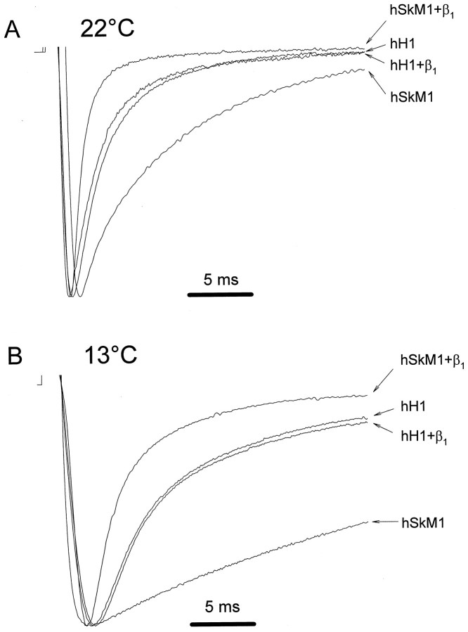
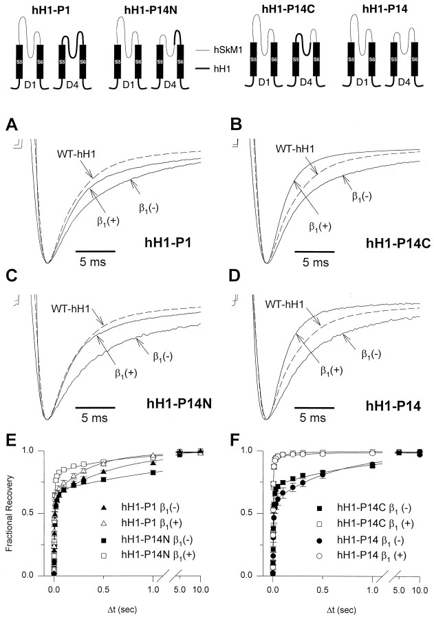
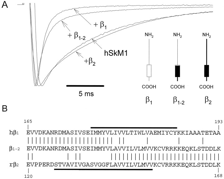
Recombinant brain, skeletal muscle, and heart voltage-gated Na+ channel alpha subunits differ in their functional responses to an accessory beta 1 subunit when coexpressed in Xenopus oocytes. We exploited the distinct beta 1 subunit responses observed for the human heart (hH1) and human skeletal muscle (hSkM1) isoforms to identify determinants of this response. Chimeric alpha subunits were constructed by exchanging the S5-S6 interhelical loops of each domain between hH1 and hSkM1 and then examined for effects on inactivation induced by coexpressed beta 1 subunit in oocytes. Substitution of single S5-S6 loops in either domain 1 (D1/S5-S6) or domain 4 (D4/S5-S6) of hSkM1 by the corresponding segments of hH1 produced channels that exhibited an attenuated response to coexpressed beta 1 subunit. Substitutions of both D1/S5-S6 and D4/S5-S6 in hSkM1 by the corresponding loops from hH1 completely abolished the effects of the beta 1 subunit on inactivation. The reciprocal chimera in which both D1/S5-S6 and D4/S5-S6 from hSkM1 were transplanted into hH1 exhibited significant beta 1 responsiveness (accelerated inactivation). The region within D4/S5-S6 that conferred beta 1 responsiveness was determined to reside primarily within an extracellular loop between the putative pore-forming segment SS2 and D4/S6. Gating modulation was also demonstrated using a chimeric beta subunit consisting of the extracellular domains of beta 1 and the transmembrane and C-terminal domains of the rat brain beta 2 subunit. These results suggest that the D1/S5-S6 and D4/S5-S6 loops in the alpha subunit and the extracellular domain of the beta 1 subunit are important determinants of the beta 1 subunit-induced gating modulation in Na+ channels.


Figures

Fig. 1.
Fig. 1.
Effect of coexpressed hβ1 on hSkM1 and hH1 gating. A, Sodium currents were recorded inXenopus oocytes expressing either wild-type hSkM1 or hH1 in the presence or absence of hβ1. Representative current traces obtained during 50 msec test depolarizations to −20 mV from a holding potential of −120 mV at room temperature (22°C) are shown. Current amplitudes are scaled to unity in all traces. Horizontal bar indicates 5 msec. B, Same experiment except that the temperature was 13°C.
Fig. 2.
Fig. 2.
Responses of wild-type and chimeric Na+ channels to coexpressed β1 subunit. Representative current tracings were recorded in the presence (asterisks) or absence (open circles) of coexpressed hβ1 during a voltage step to −20 mV from a holding potential of −120 mV. Current amplitudes are scaled and superimposed on the same time axis to illustrate the response of each channel to coexpressed β1 subunit. The composition of the α subunit chimeras constructed from hSkM1 and hH1 is illustrated next to each tracing. Filled boxes and thick lines represent structures from hH1, and open boxes and thin lines indicate hSkM1 segments. In the hH1 chimeras, vertical arrows point to the transferred hSkM1 sequences. Current decays of all the hSkM1 background chimeras except for hSkM1–P14 were significantly accelerated by coexpressed hβ1, subunit, and the hβ1subunit accelerated the inactivation kinetics of chimera hH1–P14.
Fig. 3.
Fig. 3.
Effect of coexpressed hβ1 on hSkM1–P14 or hH1–P14 Na+ channel chimeras.A, Representative current tracings illustrating failure of hβ1 to modulate inactivation in chimera hSkM1–P14. The response of hSkM1 to hβ1 in the same experiment is also shown. B, Representative current tracings illustrating accelerated time course of inactivation caused by coexpression of hβ1 with chimera hH1–P14.C, Plot of fractional recovery versus interpulse interval duration for hSkM1 (filled circles), hSkM1 + hβ1 (open circles), hSkM1–P14 (filled diamonds), and hSkM1–P14+hβ1 (open diamonds).D, Plot of fractional recovery versus interpulse interval duration (Δt) for hH1 (filled triangles), hH1–P14 (filled squares), and hH1–P14 + hβ1 (open squares). Recovery from inactivation in hH1+hβ1 is not shown but is identical to hH1 alone. (Results are represented for at least four cells; error bars are smaller than some data symbols.) Peak current amplitudes inA and B are scaled to unity. Recovery data in C and D are normalized to the current amplitude recorded after a recovery interval of 10 sec.
Fig. 4.
Fig. 4.
Sublocalization of the β1 subunit response element in D4/S5–S6. Chimeras used to sublocalize the β1 response structure in D4/S5–S6 (hH1–P14N, hH1–P14C) are illustrated by the drawings at the top of the figure. Drawings were simplified by showing only the S5–S6 region of D1 and D4 for each chimera. Structures from hSkM1 are indicated bythin lines, and segments from hH1 are indicated bythick lines or filled rectangles.AD, Representative current recordings from oocytes expressing hH1–P1 (A), hH1–P14N (B), hH1–P14C (C), or hH1–P14 (D) in the presence (+) or absence (−) of hβ1. Current sweeps of wild-type hH1 (WT–hH1, dashed lines) are provided as a reference. There were no statistical differences between time constants for hH1–P1 + β1 versus hH1–P14N + β1 and between hH1–P14C + β1 versus hH1–P14 + β1. Currents were recorded using the pulse protocol described in the legend for Figure 1, and their peak currents were scaled to unity. A representative tracing from oocytes expressing wild-type hH1 in the absence of hβ1 is shown in each figure part for comparison. E, F, Recovery from inactivation was determined as described in the legend for Figure 3 and plotted against a loga- rithmic time scale.E, Recovery curves for hH1–P14N in the presence (open squares) or absence (closed squares) of hβ1 are shown with data obtained from the hH1–P1 chimera in the presence (open triangles) or absence (closed triangles) of hβ1.F, Recovery curves for hH1–P14C in the presence (open squares) or absence (closed squares) of hβ1 are shown with data obtained from the hH1–P14 chimera in the presence (open circles) and absence (closed circles) of hβ1.
Fig. 5.
Fig. 5.
Amino acid sequence alignment of the D4/S5–S6 region. Alignment of amino acid sequences within the D4/S5–S6 region from hSkM1 (George et al., 1992), rat skeletal muscle Na+ channel μI (Trimmer et al., 1989), rat brain II Na+ channel (Auld et al., 1988), and hH1 (Gellens et al., 1992). Numbers indicate amino acid position at the beginning of each line. Residues identical to hSkM1 are shown asdashes in the aligned sequences, and the location of SS2 and D4/S6 segments is indicated by the thin lines. The β1 subunit response element is indicated by athick line. Residues within the hH1 sequence that differ from those in a consensus sequence of hSkM1, μI, and rat brain II Na+ channels are indicated byasterisks.
Fig. 6.
Fig. 6.
Effect of a β subunit chimera on hSkM1 gating.A, Scaled current recordings obtained from oocytes expressing hSkM1 alone or in the presence of wild-type hβ1 (+β1), wild-type rβ2(+β2), or a chimeric β12subunit (+β1–2). Recording conditions were the same as in Figure 1A. The figure in theinset depicts the proposed membrane topology of wild-type and chimeric β subunits. Thin lines andopen boxes indicate structures from hβ1, whereas thick black lines and filled boxes represent structures from rβ2.B, Alignment of partial amino acid sequences of hβ1, rβ2, and chimeric β1–2. The location of putative transmembrane domains of hβ1 and rβ2 is depicted by thick lines.

References

    1. Auld VJ, Goldin AL, Krafte DS, Marshall J, Dunn JM, Catterall WA, Lester HA, Davidson N, Dunn RJ. A rat brain Na+ channel α subunit with novel gating properties. Neuron. 1988;1:449–461. - PubMed
    1. Backx PH, Yue DT, Lawrence JH, Marban E, Tomaselli GF. Molecular localization of an ion-binding site within the pore of mammalian sodium channels. Science. 1992;257:248–251. - PubMed
    1. Beneski DA, Catterall WA. Covalent labeling of protein components of the sodium channel with a photactivable derivative of scorpion toxin. Proc Natl Acad Sci USA. 1980;77:639–643. - PMC - PubMed
    1. Bennett PB, Makita N, George AL., Jr A molecular basis for gating mode transitions in human skeletal muscle sodium channels. FEBS Lett. 1993;326:21–24. - PubMed
    1. Cannon SC, McClatchey AI, Gusella JF. Modification of the Na+ current conducted by the rat skeletal muscle α subunit by coexpression with a human brain β subunit. Pflügers Arch. 1993;423:155–157. - PubMed

Publication types

Substances

LinkOut - more resources