. 1996 Nov 15;16(22):7151-60.
doi: 10.1523/JNEUROSCI.16-22-07151.1996.

Physiological mapping of local inhibitory inputs to the hypothalamic paraventricular nucleus

Affiliations

Physiological mapping of local inhibitory inputs to the hypothalamic paraventricular nucleus

C Boudaba et al. J Neurosci. .

Abstract

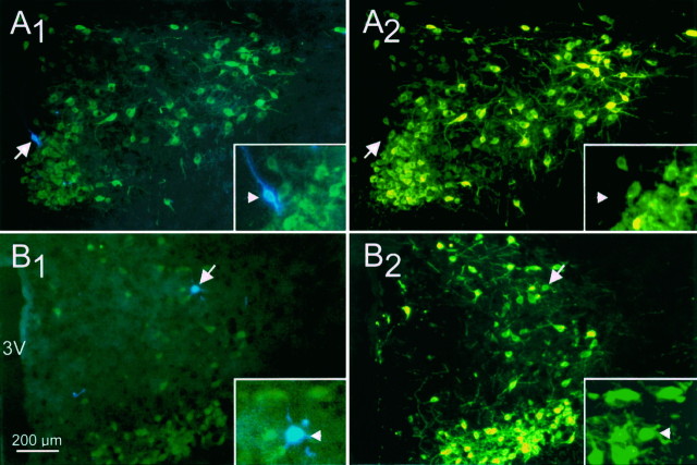
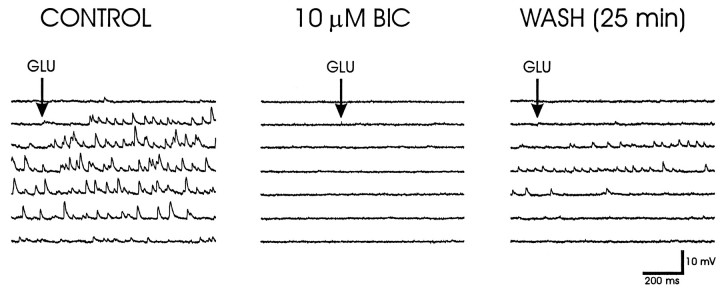
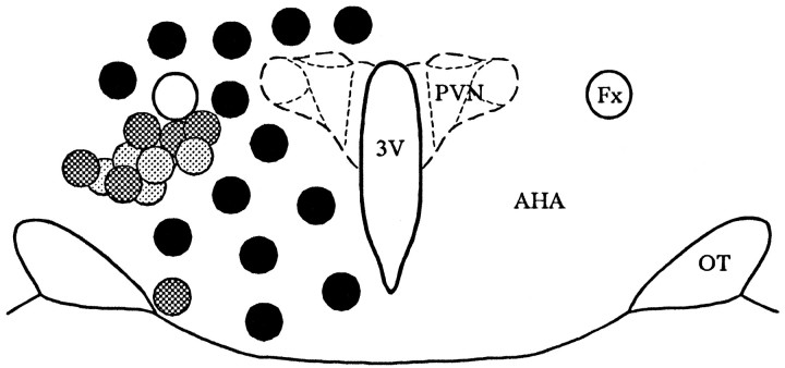
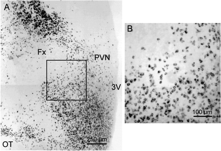
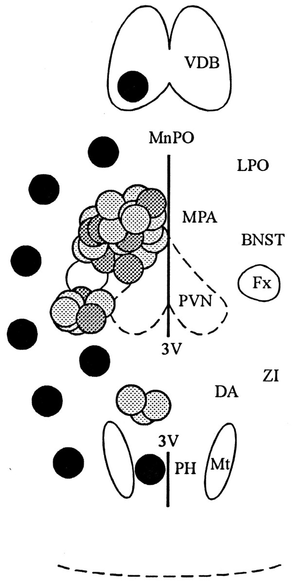
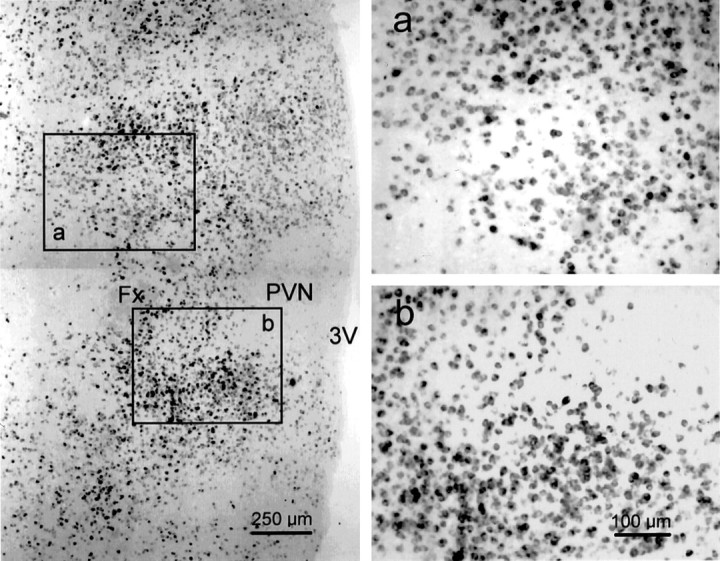
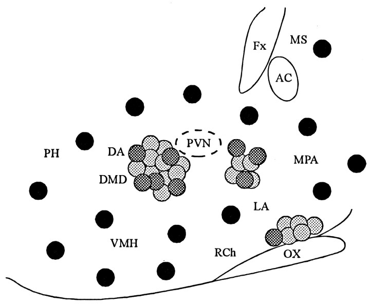
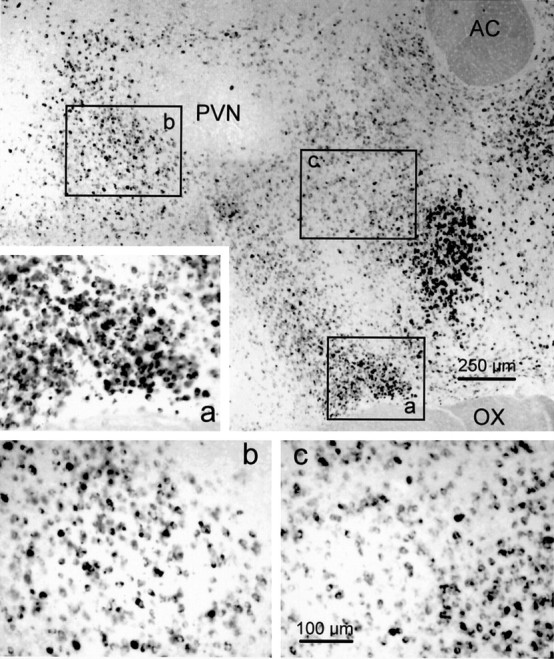
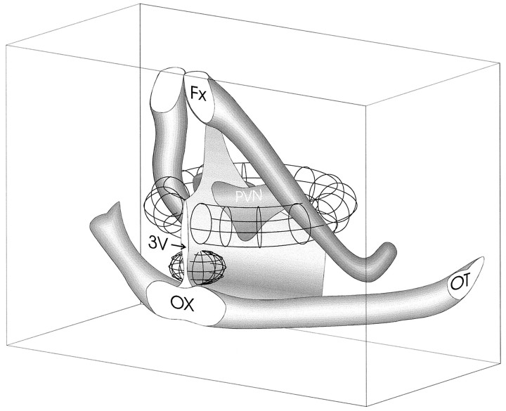
Local inhibitory synaptic inputs to neurons of the rat hypothalamic paraventricular nucleus (PVN) were studied by using glutamate microstimulation and conventional intracellular and whole-cell patch-clamp recording in coronal, horizontal, and parasagittal slices of rat hypothalamus. PVN cells were classified as magnocellular or parvocellular neurons on the basis of electrophysiological and post hoc immunohistochemical analyses; GABA-producing neurons were localized with in situ hybridization. Glutamate microstimulation of different sites around the PVN evoked volleys of postsynaptic potentials in 43% of the PVN neurons tested. Some responses to stimulation at each site were blocked by bicuculline, suggesting that they were mediated by the activation of presynaptic GABA neurons. In the coronal plane, presynaptic inhibitory sites were located lateral to the PVN and ventral to the fornix, corresponding to the lateral hypothalamic area and the posterior bed nucleus of the stria terminalis (BNST). In the horizontal plane, presynaptic inhibitory sites were found rostral, lateral, and caudal to the nucleus, corresponding to parts of the anterior hypothalamic area, the posterior BNST, the medial preoptic area, and the dorsomedial hypothalamus. In the parasagittal plane, presynaptic inhibitory neurons were revealed at sites rostral and caudal to the nucleus, corresponding to the medial preoptic area and the dorsomedial hypothalamus, and in a site dorsal to the optic chiasm that included the suprachiasmatic nucleus. These presynaptic sites each contained GABA-producing neurons based on in situ hybridization with a glutamic acid decarboxylase riboprobe and together formed a three-dimensional ring around the PVN. Unexpectedly, both magnocellular and parvocellular neurons received inhibitory synaptic inputs from common sites.


Figures

Fig. 1.
Fig. 1.
Immunohistochemical identification of neurons recorded in the PVN. Biocytin-labeled cells were visualized under the blue AMCA filter combination (A1, B1), and cells stained immunohistochemically were examined under fluorescein filters (A2, B2). The FITC fluorescence can be detected with the blue filter combination, but the AMCA is not detectable under the fluorescein filters. A, A PVN cell classified as a putative parvocellular neuron on the basis of its electrical properties was labeled with the intracellular biocytin/AMCA marker (A1); this cell was negative for the FITC neurophysin immunolabel (A2), confirming that it was a parvocellular neuron. B, A PVN cell classified electrophysiologically as a magnocellular neuron was double-labeled with the intracellular biocytin/AMCA label (B1) and with the FITC immunolabel for oxytocin (B2), indicating that it was an oxytocinergic magnocellular neuron. Sections in A andB were taken from horizontal slices.Insets show the areas containing the biocytin-labeled cells (arrows) enlarged twofold. 3V, Third ventricle.
Fig. 2.
Fig. 2.
Glutamate-evoked IPSPs. Left, Glutamate microstimulation (GLU) at a position ventral to the fornix elicited reversed IPSPs in a PVN parvocellular neuron recorded with a KCl-filled microelectrode.Middle, Bath application of the GABAA-receptor antagonist bicuculline methiodide (BIC) for 15 min completely blocked the effect of glutamate microapplication at the same site and with the same application parameters. Right, Partial recovery of the glutamate-evoked IPSPs was seen after 25 min of washout of the bicuculline. The membrane potential was held at −105 mV with negative current injection.
Fig. 3.
Fig. 3.
Topographic distribution of presynaptic active zones in the coronal slice. Glutamate microstimulation at positions lateral to the PVN and ventral to the fornix elicited an increase in PSPs recorded in PVN neurons, shown with light anddark gray circles. Each gray circlerepresents the site at which glutamate microstimulation elicited a synaptic response in a PVN cell. The dark gray circlesrepresent the sites at which the glutamate-evoked synaptic responses were verified as inhibitory (see text). The microstimulation sites at which no synaptic response was recorded in PVN neurons are shown withblack circles. The schematic diagram of the hypothalamus was modified from the atlas of Swanson (1992). AHA, Anterior hypothalamic area; Fx, fornix;OT, optic tract; 3V, third ventricle.
Fig. 4.
Fig. 4.
Distribution of GAD65 mRNA-containing neurons in the coronal plane of the hypothalamus. A, Low-magnification photomontage of a coronal section labeled with the GAD65 riboprobe. GAD65 mRNA was expressed in neurons concentrated in regions dorsal to the fornix (Fx), corresponding to the zona incerta, and ventrolateral to the PVN in the anterior hypothalamic area. Some weakly labeled neurons can be seen within the PVN, although a greater density of labeled cells was found around the ventrolateral edge of the nucleus. The hypothalamic region delineated by thebox represents part of the area ventrolateral to the PVN in which glutamate microstimulation was effective at eliciting IPSPs in PVN neurons; it is shown at higher magnification in B.OT, Optic tract; 3V, third ventricle.
Fig. 5.
Fig. 5.
Topographic distribution of presynaptic active zones in the horizontal slice. Glutamate microstimulation elicited PSPs in PVN neurons when applied in three positions in the horizontal plane: rostral to the PVN and to the fornix, lateral to the PVN and caudal to the fornix, and caudal to the PVN, as shown with lightand dark gray circles. Each gray circlerepresents the site at which glutamate microstimulation elicited a synaptic response in a PVN neuron. Sites at which glutamate microstimulation evoked synaptic responses that were confirmed as inhibitory are shown with dark gray circles. No synaptic responses were recorded with glutamate microstimulation in the locations shown with black circles. Rostral isup in this diagram. BNST, Bed nucleus of the stria terminalis; Fx, fornix; DA, dorsal hypothalamic area; LPO, lateral preoptic area;MnPO, median preoptic nucleus; MPA, medial preoptic area; Mt, mamillothalamic tract;PH, posterior hypothalamic area; 3V, third ventricle; VDB, vertical limb of the diagonal band of Broca; ZI, zona incerta.
Fig. 6.
Fig. 6.
Distribution of GAD65 mRNA-containing neurons in the horizontal plane of the hypothalamus. Low-magnification photomontage of a horizontal section labeled with the GAD65 riboprobe (left). GAD65 mRNA is expressed in neurons scattered throughout the hypothalamus, including cells surrounding the PVN, which itself is relatively devoid of GAD65 mRNA-containing cells. The sites rostral and caudal to the fornix that were stimulated by glutamate microdrops to elicit IPSPs in PVN neurons are represented byboxes a and b, respectively, which are shown at higher magnification to the right. The midline is to the right, and rostral is up in each photomicrograph. The calibration in b pertains to both a and b. Fx, Fornix;3V, third ventricle.
Fig. 7.
Fig. 7.
Topographic distribution of presynaptic active zones in the parasagittal slice. PSPs were evoked in PVN neurons with glutamate microstimulation in three positions in the parasagittal plane: caudal to the PVN, rostral to the PVN, and at the base of the hypothalamus dorsal to the optic chiasm, as shown by thelight and dark gray circles. Eachgray circle marks the spot at which glutamate microstimulation evoked a synaptic response in a PVN neuron. The sites at which the glutamate microdrops elicited synaptic responses identified as IPSPs are shown by the dark gray circles. The positions in which glutamate microdrops never elicited a synaptic response are designated with black circles. Rostral is to the right. AC, Anterior commissure;DA, dorsal hypothalamic area; DMD, dorsomedial hypothalamic nucleus; Fx, fornix;LA, lateral hypothalamus; MPA, medial preoptic area; MS, medial septum; OX, optic chiasm; PH, posterior hypothalamic area;Rch, retrochiasmatic area; VMH, ventromedial hypothalamic nucleus.
Fig. 8.
Fig. 8.
Distribution of GAD65 mRNA-containing cells in the parasagittal plane of the hypothalamus. Shown is a photomontage at low magnification of a parasagittal section labeled with the GAD65 riboprobe. GAD65 mRNA was expressed in cells concentrated most heavily in areas rostral to the PVN, corresponding to the medial preoptic area and dorsal to the optic chiasm (OX), including the suprachiasmatic nucleus. Inset a, shown at higher magnification to the left, represents the ventral site at which glutamate microstimulation elicited IPSPs in PVN neurons recorded in parasagittal slices. This site had a high density of strongly labeled neurons. The other two sites responsive to glutamate microstimulation caudal and ventrorostral to the PVN are designated byboxes b and c, respectively; these sites contained a qualitatively moderate density of GAD65 mRNA-expressing neurons and are shown at higher magnification below(b, c). The second site (box b) is located in the dorsomedial hypothalamic area. The third site (box c) includes the anterior hypothalamic and medial preoptic areas, the striohypothalamic nucleus, and the medial part of the posterior bed nucleus of the stria terminalis. Rostral is to the right. AC, Anterior commissure;OX, optic chiasm.
Fig. 9.
Fig. 9.
Three-dimensional rendering of presynaptic active zones. The topographic maps of presynaptic active sites in the coronal, horizontal, and parasagittal planes were rendered to create a qualitative schematic diagram depicting an approximated three-dimensional topography of zones concentrating PVN-projecting GABA neurons. The lateral sites together form a ring that courses ventrally and laterally around the PVN, and the ventral zone is situated just dorsal to the optic chiasm. The lateral spread of the ventral zone is unknown, because only the parasagittal plane that included the PVN was tested. Fx, Fornix; OT, optic tract;OX, optic chiasm; 3V, third ventricle.

References

    1. Altstein M, Whitnall MH, House S, Key S, Gainer H. An immunochemical analysis of oxytocin and vasopressin prohormone processing in vivo. Peptides. 1988;9:87–105. - PubMed
    1. Arluison M, Brochier G, Vankova M, Leviel V, Villalobos J, Tramu G. Demonstration of peptidergic afferents to the bed nucleus of the stria terminalis using local injections of colchicine. A combined immunohistochemical and retrograde tracing study. Brain Res Bull. 1994;34:319–337. - PubMed
    1. Boudaba C, Poulain DA. Further evidence that the septum is not part of the main pathway of the milk ejection in the rat. J Neuroendocrinol. 1991;3:199–204. - PubMed
    1. Boudaba C, Szabó K, Tasker JG. Physiological mapping of local inhibitory neurons which project to the paraventricular nucleus in rat hypothalamic slices. Soc Neurosci Abstr. 1995;21:1665.
    1. Bourque CW. Transient calcium-dependent potassium current in the magnocellular neurosecretory cells of the rat supraoptic nucleus. J Physiol (Lond) 1988;397:331–347. - PMC - PubMed

Publication types

LinkOut - more resources