. 1996 Nov 15;16(22):7240-52.
doi: 10.1523/JNEUROSCI.16-22-07240.1996.

A role in migration for the alpha V beta 1 integrin expressed on oligodendrocyte precursors

Affiliations

A role in migration for the alpha V beta 1 integrin expressed on oligodendrocyte precursors

R Milner et al. J Neurosci. .

Abstract

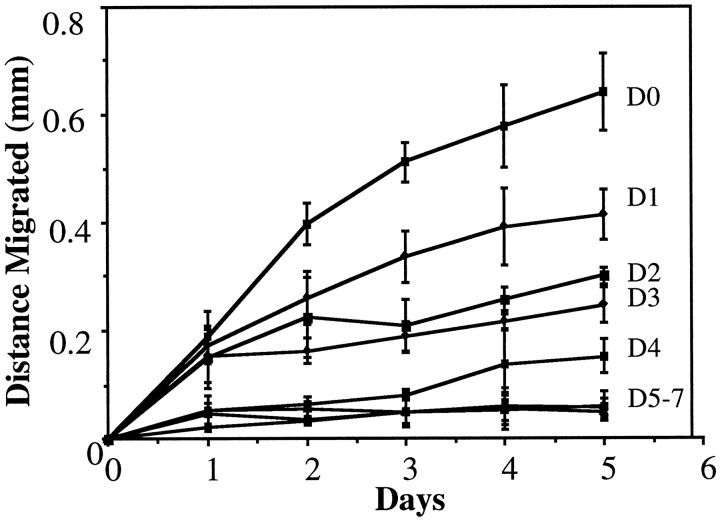
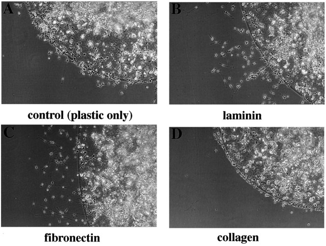
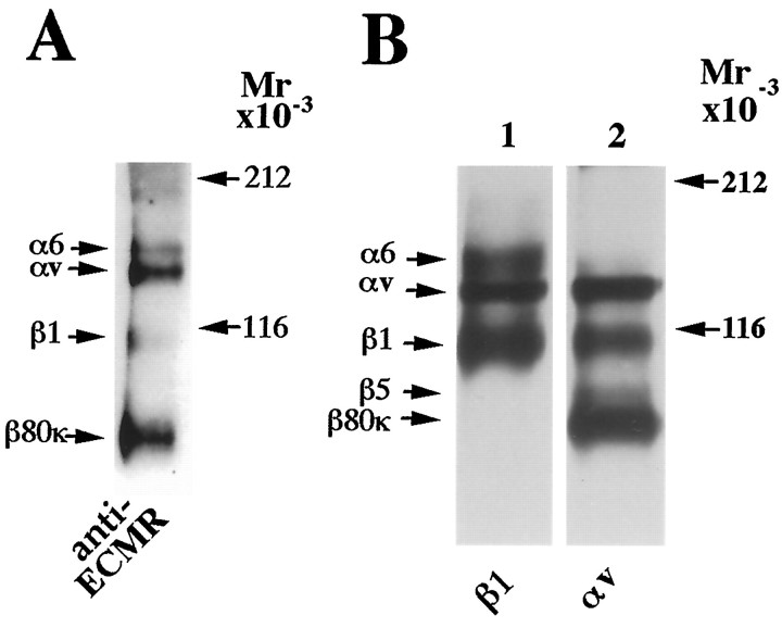
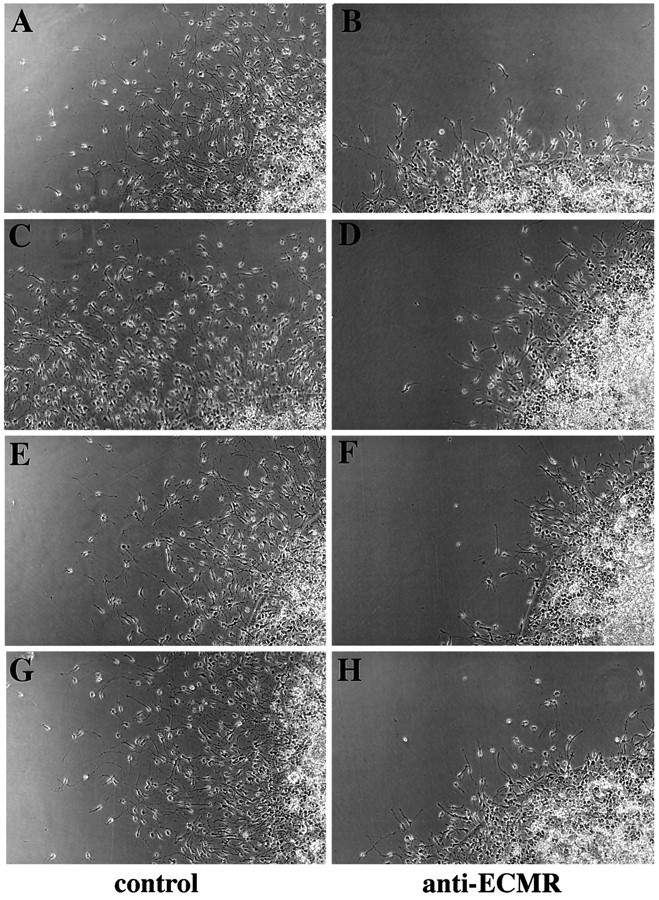
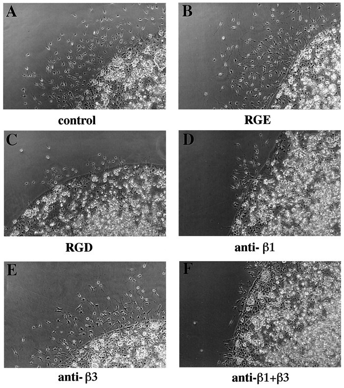
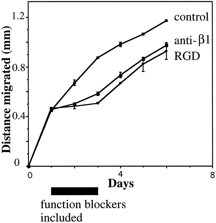
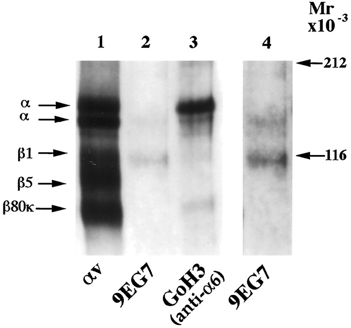
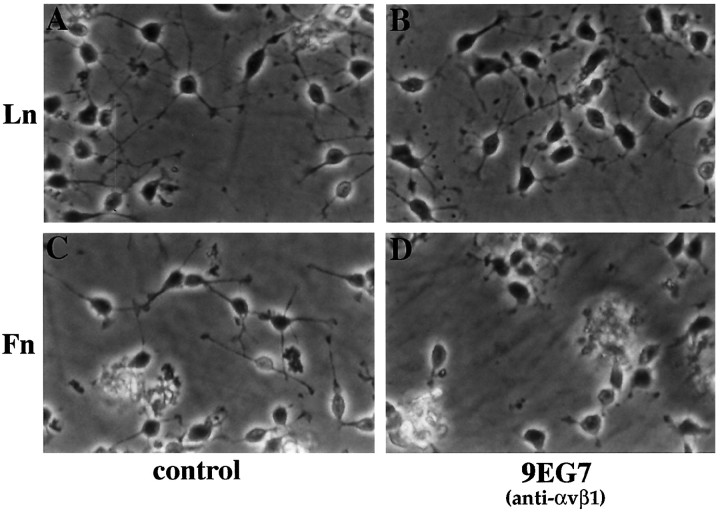
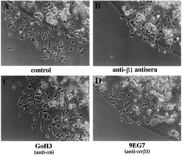
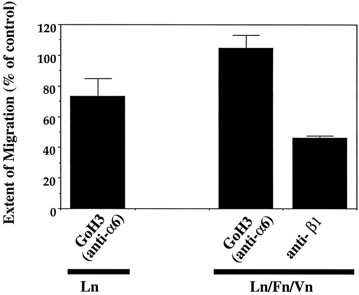
Myelination of the CNS requires the migration of oligodendrocyte precursors throughout the CNS from restricted regions within the ventricular and subventricular zones. In light of the significant effects of cell-extracellular matrix (ECM) interactions on cell migration in other developing systems, we have analyzed the role of integrins in oligodendrocyte precursor migration. We have shown previously that oligodendrocyte precursors in vitro express a limited repertoire of integrins, including alpha 6 beta 1, alpha v beta 3, and that differentiation is associated with downregulation of alpha v beta 1 and upregulation of alpha v beta 5. Using a migration assay based on the movement of cells away from an agarose drop containing a high-density cell suspension, we find that RGD peptides (that block alpha v but not alpha 6 integrins) and anti-beta 1 antibodies block migration on an astrocyte-derived ECM, whereas anti-beta 3 antibodies have little effect. These results suggest that alpha v beta 1 but not alpha 6 beta 1 plays a role in oligodendrocyte precursor migration, and this is confirmed by the use of blocking monoclonal antibodies that distinguish these two integrins. In keeping with the results of others, we find that differentiated oligodendrocytes lose migratory potential and that the timing of this loss correlates with downregulation of alpha v beta 1. Taken together with the work of others showing that ECM ligands for alpha v beta 1 are expressed within the CNS, we propose that this integrin plays a significant role in the migration of oligodendrocyte precursors in vivo and that its downregulation during differentiation could be an important factor regulating the migratory phenotype of these cells.


Figures

Fig. 1.
Fig. 1.
The effect of PDGF on the migration of oligodendrocyte precursors. Cells were resuspended at high density in agarose, as described in Materials and Methods, and then plated as small drops onto poly-dl-ornithine-coated plastic and cultured in the absence (A) or presence (B) of PDGF (5 ng/ml). Note that PDGF promotes the migration of oligodendrocyte precursors, with no migration observed in the absence of this growth factor.
Fig. 2.
Fig. 2.
The relationship between differentiation and migration in oligodendrocyte precursor cells. Cells were resuspended at high density in agarose, as described in Materials and Methods, and then plated as small drops onto poly-dl-ornithine-coated plastic and grown in defined medium alone, without PDGF. Cells were then allowed to differentiate for 0–7 d before PDGF was added to promote migration, and migration was measured for the following 5 d. Data from three experiments are shown. Note that the more the cell populations are allowed to differentiate before the addition of PDGF (most differentiated at day 7, least at day 0), the less they migrate in response to PDGF.
Fig. 3.
Fig. 3.
The migration of oligodendrocyte precursors on different ECM substrates. Cells were resuspended at high density in agarose, as described in Materials and Methods, and then plated as small drops onto uncoated tissue-culture plastic. The ECM substrate was then added, as described in Materials and Methods. A, Control with no ECM; B, laminin; C, fibronectin; D, collagen. Note that after 2 d, cells on laminin and fibronectin had migrated further than the uncoated plastic control. Cells on collagen, in contrast, had migrated less than the control and have lined up around the periphery of the agarose drop.
Fig. 4.
Fig. 4.
Immunoprecipitations of integrins from rat oligodendrocyte precursors to illustrate the specificity of the polyclonal antibodies used. Immunoprecipitations of biotin-labeled cell-surface proteins were performed, as described in Materials and Methods, with the anti-ECMR antiserum (A), anti-β1 antiserum (B, lane 1), or anti-αv antisera (B, lane 2). After this, the proteins were separated on nonreducing gels. Note that the anti-ECMR antiserum immunoprecipitates a repertoire of bands corresponding to all of the integrins expressed by oligodendrocyte precursors (α6β1, αvβ1, and αvβ80k). The anti-β1 antiserum recognizes the β1 subunit in association with two α subunits corresponding to α6 and αv, but it does not cross-react with β80k.
Fig. 5.
Fig. 5.
Effect of the anti-ECMR antiserum on oligodendrocyte precursor migration on different substrates. Cell migration away from agarose drops (prepared as described in Materials and Methods) after 2 d on poly-dl-ornithine (A, B), laminin (C, D), fibronectin (E, F), or vitronectin (G, H) is shown in the presence of either normal goat serum (A, C, E, G) or anti-ECMR antiserum (B, D, F, H), both at 1:400. Note that the anti-ECMR antiserum reduces oligodendrocyte precursor migration on all substrates tested but does not alter the bipolar morphology of the cells.
Fig. 6.
Fig. 6.
The effect of integrin inhibitors on oligodendrocyte precursor migration on AGM. Cell migration away from agarose drops (prepared as described in Materials and Methods) after 2 d on AGM is shown in the presence of normal rabbit serum (A), RGE peptide (B), RGD peptide (C), anti-β1 antiserum (D), anti-αvβ3 antiserum (E), or anti-β1 and anti-αvβ3 antiserum (F). The peptides were present at 0.1 mg/ml. Note that cell migration was inhibited both by RGD peptides and anti-β1 antiserum without any change to the morphology of the cells.
Fig. 7.
Fig. 7.
Quantification of the effect of integrin inhibitors on oligodendrocyte precursor migration on AGM. Cell migration away from agarose drops (prepared as described in Materials and Methods) after 2 d on AGM was measured in the presence of either normal rabbit serum, RGE peptide, RGD peptide, anti-β1 antiserum, anti-αvβ3 antiserum, F11 (monoclonal anti-β3), or anti-β1 and anti-αvβ3 antiserum in combination. The peptides were present at 0.1 mg/ml. Each point represents the mean ± SEM of three separate experiments. Note that cell migration was inhibited only by RGD peptides and anti-β1 antiserum.
Fig. 8.
Fig. 8.
Migration of oligodendrocyte precursor cells after removal of integrin blockade. Cells were allowed to migrate away from agarose drops (prepared as described in Materials and Methods) for 1 d before the following reagents were introduced: RGE peptide (control), RGD peptide, or anti-β1 antiserum. After incubation for 2 d, the reagents were removed and cell migration was measured for an additional 3 d. Note that cells whose migration was blocked with either the RGD peptide or anti-β1 antiserum migrate at equal rates to the control cells once the blockade is removed.
Fig. 9.
Fig. 9.
Specificity of the 9EG7 monoclonal antibody for oligodendroglial αvβ1 integrin. Immunoprecipitations of biotin-labeled cell-surface proteins from mouse oligodendrocyte precursors were performed as described in Materials and Methods, with either anti-αv antiserum (lane 1), the 9EG7 monoclonal antibody (lane 2), or the anti-α6-specific GoH3 monoclonal antibody (lane 3). The proteins were separated on a nonreducing gel. Lanes 1–3 represent three adjacent lanes on the gel that have been exposed for equal lengths of time, whereas lane 4 represents a longer exposure of lane 2. Note that the 9EG7 antibody immunoprecipitates the β1 subunit associated with an α subunit that comigrates with the dominant αv subunit but not the α6 subunit.
Fig. 10.
Fig. 10.
Specific function-blocking effect of the anti-αvβ1 9EG7 monoclonal antibody. Mouse oligodendrocyte precursors were plated onto either laminin (A, B) or fibronectin (C, D) in the absence (A, C) or presence (B, D) of the 9EG7 monoclonal antibody, and allowed to adhere and process for 1 hr. Note that 9EG7 inhibits process outgrowth on fibronectin but not laminin.
Fig. 11.
Fig. 11.
The effect of integrin inhibitors on oligodendrocyte precursor migration on AGM. Mouse oligodendrocyte precursor cell migration away from agarose drops (prepared as described in Materials and Methods) after 2 d on AGM was measured under control conditions (A) or in the presence of anti-β1 antiserum (B), the anti-α6 GoH3 monoclonal antibody (C), or the anti-αvβ1 9EG7 monoclonal antibody (D). Note that cell migration was inhibited by the anti-β1 antiserum and the anti-αvβ1 9EG7 monoclonal antibody but not by the anti-α6 GoH3 antibody.
Fig. 12.
Fig. 12.
The effect of αvβ1 and α6β1 inhibition on oligodendrocyte precursor migration over ECM substrates. The extent of mouse oligodendrocyte precursor cell migration away from agarose drops (prepared as described in Materials and Methods) after 2 d on either laminin or a composite laminin/fibronectin/vitronectin substrate was measured in the presence of the anti-α6 GoH3 monoclonal antibody or anti-β1 antiserum. The extent of migration is presented as a percentage of migration observed under control conditions, with no antibody present. Note that the anti-α6 GoH3 antibody had no significant inhibitory effect on either laminin or the composite substrate (p > 0.05), whereas the anti-β1 antiserum significantly inhibited migration on the composite substrate (p < 0.001).

Similar articles

Cited by

References

    1. Adams JC, Watt FM. Regulation of development and differentiation by the extracellular matrix. Development. 1993;117:1183–1198. - PubMed
    1. Albelda SM, Mette SA, Elder DE, Stewart R, Damjanovich L, Herlyn M, Buck C. Integrin distribution in malignant melanoma: association of the beta 3 subunit with tumour progression. Cancer Res. 1990;50:6757–6764. - PubMed
    1. Alfandari D, Whittaker CA, DeSimone DW, Darribere T. Integrin αv subunit is expressed on mesodermal cell surfaces during amphibian gastrulation. Dev Biol. 1995;170:249–261. - PubMed
    1. Almeida EAC, Huovila A-PJ, Sutherland AE, Stephens LE, Calarco PG, Shaw LM, Mercurio AM, Sonnenberg A, Primakoff P, Myles DG, White JM. Mouse egg integrin α6β1 functions as a sperm receptor. Cell. 1995;81:1095–1104. - PubMed
    1. Armstrong RC, Harvath L, Dubois-Dalcq ME. Type 1 astrocytes and oligodendrocyte-type 2 astrocyte glial progenitors migrate toward distinct molecules. J Neurosci Res. 1990;27:400–407. - PubMed

Publication types

MeSH terms

LinkOut - more resources