. 1996 Nov 15;16(22):7308-17.
doi: 10.1523/JNEUROSCI.16-22-07308.1996.

An explanation for reflex blink hyperexcitability in Parkinson's disease. I. Superior colliculus

Affiliations

An explanation for reflex blink hyperexcitability in Parkinson's disease. I. Superior colliculus

M A Basso et al. J Neurosci. .

Abstract

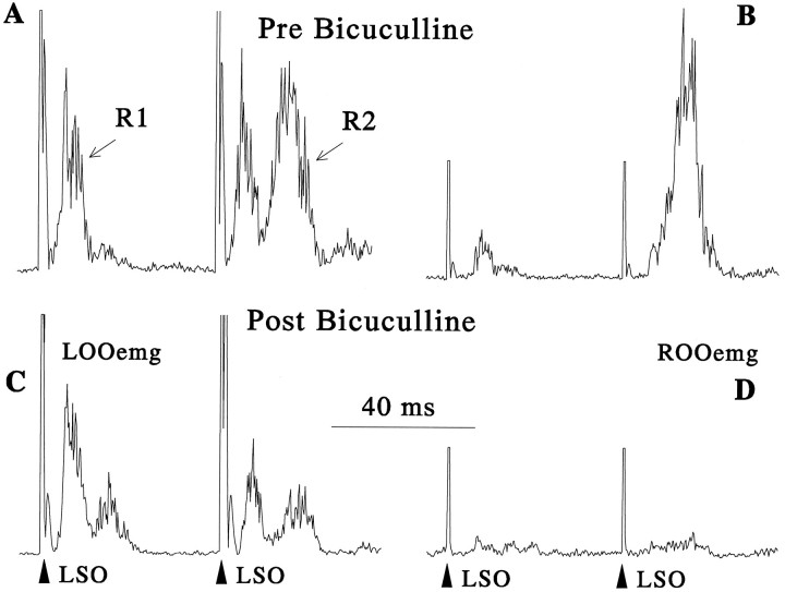
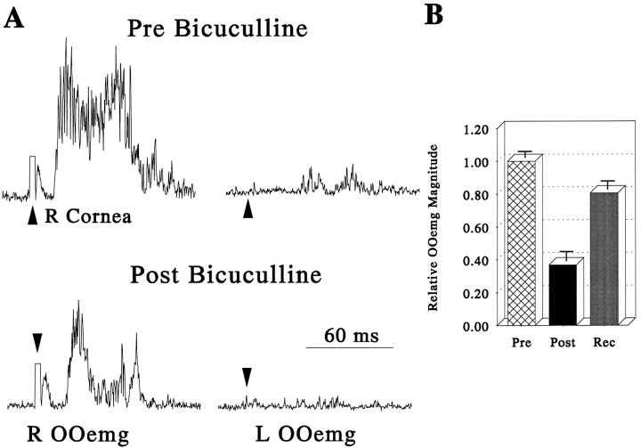
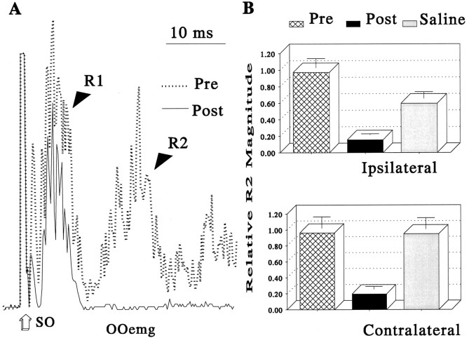
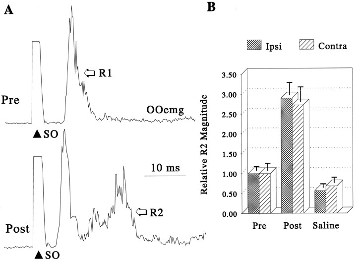
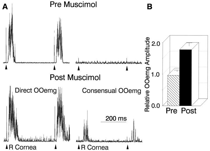
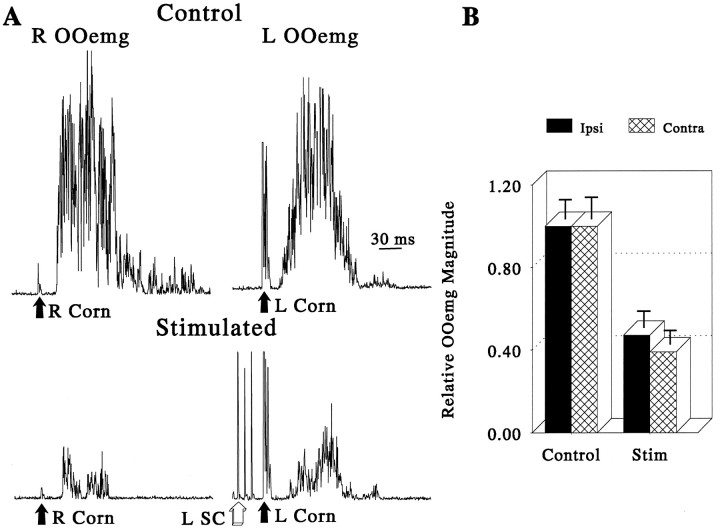
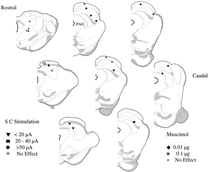
Hyperexcitable reflex blinks are a cardinal sign of Parkinson's disease. We investigated the neural circuit through which a loss of dopamine in the substantia nigra pars compacta (SNc) leads to increased reflex blink excitability. Through its inhibitory inputs to the thalamus, the basal ganglia could modulate the brainstem reflex blink circuits via descending cortical projections. Alternatively, with its inhibitory input to the superior colliculus, the basal ganglia could regulate brainstem reflex blink circuits via tecto-reticular projections. Our study demonstrated that the basal ganglia utilizes its GABAergic input to the superior colliculus to modulate reflex blinks. In rats with previous unilateral 6-hydroxydopamine (6-OHDA) lesions of the dopamine neurons of the SNc, we found that microinjections of bicuculline, a GABA antagonist, into the superior colliculus of both alert and anesthetized rats eliminated the reflex blink hyperexcitability associated with dopamine depletion. In normal, alert rats, decreasing the basal ganglia output to the superior colliculus by injecting muscimol, a GABA agonist, into the substantia nigra pars reticulata (SNr) markedly reduced blink amplitude. Finally, brief trains of microstimulation to the superior colliculus reduced blink amplitude. Histological analysis revealed that effective muscimol microinjection and microstimulation sites in the superior colliculus overlapped the nigrotectal projection from the basal ganglia. These data support models of Parkinsonian symtomatology that rely on changes in the inhibitory drive from basal ganglia output structures. Moreover, they support a model of Parkinsonian reflex blink hyper-excitability in which the SNr-SC target projection is critical.


Figures

Fig. 1.
Fig. 1.
Elimination of blink reflex hyperexcitability by a microinjection of bicuculline into the superior colliculus of an alert rat with a 6-OHDA lesion of the right ascending mesotelencephalic dopamine system. Each trace is the average of 10 rectified OOemg responses to a pair of stimuli to the left supraorbital branch of the trigeminal nerve (LSO) separated by 50 msec. A, C, The left records are from the left lid OOemg (LOOemg), the direct response, and the (B, D)right records are consensual, right lid OOemg (ROOemg) responses. A, B, The top records are before microinjection of bicuculline (Pre Bicuculline). C, D, The bottom records are after a microinjection of 1 μg of bicuculline into the right superior colliculus.LOOemg, Left orbicularis oculi response;LSO, left supraorbital nerve stimulation;R1, short-latency response; R2, long-latency response; ROOemg, right orbicularis oculi emg.
Fig. 2.
Fig. 2.
Elimination of blink reflex hyperexcitability by a microinjection of bicuculline into the superior colliculus of a urethane-anesthetized rat with a 6-OHDA lesion of the ascending mesotelencephalic dopamine system. A, Each trace is the average of 10 rectified OOemg responses to stimulation of the right cornea (R Cornea). Left traces show the right OOemg (R OOemg), the direct response, and theright traces display the left OOemg (L OOemg), the consensual response, not typically seen in normal rats. The top traces illustrate the direct and consensual response before microinjection of bicuculline (Pre Bicuculline), and the bottom traces show the same responses after microinjection of 0.5 μg of bicuculline into the superior colliculus (Post Bicuculline).B, Pooled data from three anesthetized rats with 6-OHDA lesions showing the corneally evoked OOemg amplitude before (Pre), after (Post), and at least 2 hr after (Rec) microinjections of bicuculline into the superior colliculus ipsilateral to the 6-OHDA lesion. All integrated OOemg data were normalized to the mean preinjection OOemg amplitude for all animals, and error bars are 1 SEM.
Fig. 3.
Fig. 3.
Reduction of substantia nigra pars reticulata activity by microinjection of muscimol reduced R2 OOemg amplitude in alert rats. A, Each trace is the average of 15 rectified, direct OOemg responses to stimulation of the supraorbital branch of the trigeminal nerve (SO) before (dotted line) and after (solid line) a 0.5 μl microinjection of 1.0% muscimol into the SNr.B, R2 OOemg amplitude relative to premicroinjection OOemg amplitude before (Pre) and after (Post) microinjections of muscimol and after a control microinjection of saline (Saline) from two alert rats.Top graph shows data from direct OOemg activity ipsilateral to the SNr microinjection, and bottom graphdisplays direct OOemg activity contralateral to the SNr microinjection. Error bars are 1 SEM.
Fig. 4.
Fig. 4.
Microinjection of muscimol into the superior colliculus increases R2 blink component amplitude in alert rats.A, Each record is the average of 15 rectified, direct OOemg responses to stimulation of the supraorbital branch of the trigeminal nerve (SO) before (top trace, Pre) and after (bottom trace, Post) a 0.1 μg muscimol microinjection into the superior colliculus contralateral to the SO stimulus. B, Relative R2 OOemg amplitude evoked by SO stimuli presented ipsilateral (hatched bars) or contralateral (striped bars) to the injected superior colliculus. The R2 component increased bilaterally (Post) relative to stimuli delivered before the microinjection (Pre) or after control microinjections of saline (Saline). All integrated OOemg data were normalized to the mean preinjection OOemg amplitude, and error bars are 1 SEM.
Fig. 5.
Fig. 5.
Microinjection of muscimol into the superior colliculus of urethane-anesthetized rats mimics the effects of 6-OHDA lesions. A, Each record is the average of 10 rectified OOemg responses to two stimuli delivered to the right cornea (R Cornea) with an interstimulus interval of 400 msec. Theleft traces are from the right OOemg (Direct OOemg), and right traces are from the left OOemg (Consensual OOemg). Top traces are direct and consensual blinks evoked before muscimol microinjection (Pre Muscimol), and the bottom traces are blinks evoked after a 0.01 μg microinjection of muscimol into the left superior colliculus (Post Muscimol). B, Relative direct OOemg amplitude evoked by SO stimulation before (Pre, striped bars) and after (Post, solid bars) muscimol microinjection into the SC contralateral to the corneal stimulus. The bars are means of nine microinjections normalized to the mean preinjection OOemg amplitude of all animals, and the error bars are 1 SEM.
Fig. 6.
Fig. 6.
Microstimulation of the superior colliculus suppresses cornea reflex blinks in urethane-anesthetized rats.A, Each record is the average of 10 rectified OOemg responses to stimulation of the right (R Corn) or left (L Corn) cornea. Right traces are from the left OOemg (L OOemg), and left tracesare from the right lid OOemg (R OOemg). Top traces show normal, control responses, and bottom traces illustrate responses preceded by a 70 msec train of microstimulation to the left superior colliculus (L SC) that terminated 5 msec before the corneal stimulation.B, Reduction in the amplitude of the direct OOemg response caused by microstimulation of the superior colliculus ipsilateral (Ipsi, solid bars) or contralateral (Contra, hatched bars) to the stimulated cornea. All integrated OOemg data were normalized to the mean preinjection OOemg amplitude for the group, and error bars are 1 SEM.
Fig. 7.
Fig. 7.
Reconstruction of the sites of muscimol microinjections and microstimulation sites in anesthetized rats. Microinjection and microstimulation sites from all animals mapped on schematic representation of coronal brain sections spaced 200 μm apart (Swanson, 1992). Filled diamonds are sites where 0.01 μg of muscimol increased blink amplitude; × are sites where 0.1 μg of muscimol increased blink amplitude. Inverted filled triangles show sites where currents <20 μA caused a 50% reduction in blink amplitude. Filled squares show sites where currents between 20 and 40 μA caused a 50% reduction in blink amplitude. Filled circles show sites requiring currents > 50 μA to cause a 50% reduction in blink amplitude.Filled stars are sites where 0.1 μg of muscimol did not alter reflex blinks. Open stars show sites where currents > 100 μA failed to alter reflex blinks.PAG, Periaqueductal gray; sg, stratum griseum superficiale; si, stratum griseum intermediale.

References

    1. Albin RL, Young AB, Penney JB. The functional anatomy of basal ganglia disorders. Trends Neurosci. 1989;12:366–375. - PubMed
    1. Basso MA, Evinger C. An explanation for blink reflex hyperexcitability in Parkinson’s disease. II. Nucleus raphe magnus. J Neurosci. 1996;16:7318–7330. - PMC - PubMed
    1. Basso MA, Strecker RE, Evinger C. Midbrain 6-hydroxydopamine lesions modulate blink reflex excitability. Exp Brain Res. 1993;94:88–96. - PubMed
    1. Berardelli A, Accornero N, Cruccu G, Fabiano F, Guerrisi V, Manfredi M. The orbicularis oculi response after hemispheral damage. J Neurol Neurosurg Psychiatry. 1983;46:837–843. - PMC - PubMed
    1. Bickford ME, Hall WC. The nigral projection to predorsal bundle cells in the superior colliculus of the rat. J Comp Neurol. 1992;319:11–33. - PubMed

Publication types

LinkOut - more resources