. 1996 Nov 15;16(22):7318-30.
doi: 10.1523/JNEUROSCI.16-22-07318.1996.

An explanation for reflex blink hyperexcitability in Parkinson's disease. II. Nucleus raphe magnus

Affiliations

An explanation for reflex blink hyperexcitability in Parkinson's disease. II. Nucleus raphe magnus

M A Basso et al. J Neurosci. .

Abstract

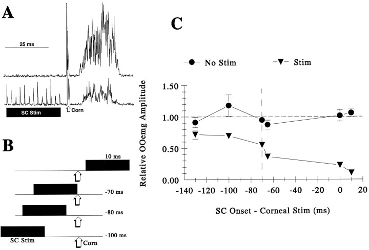
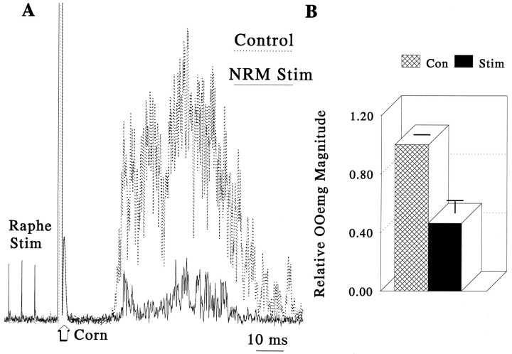
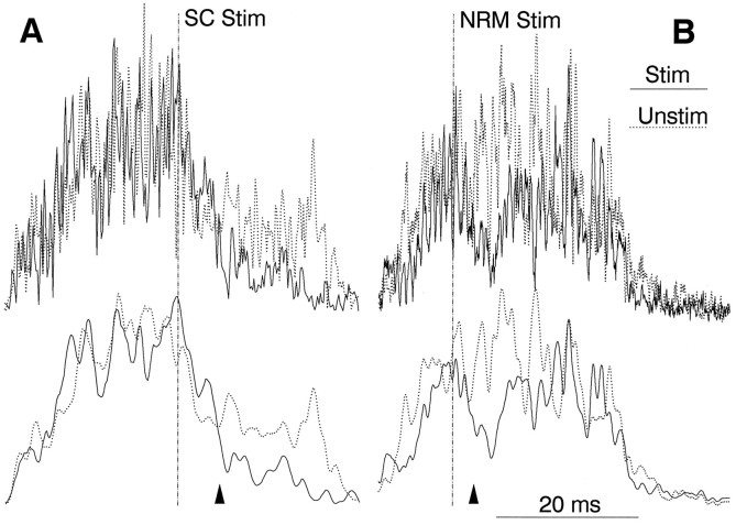
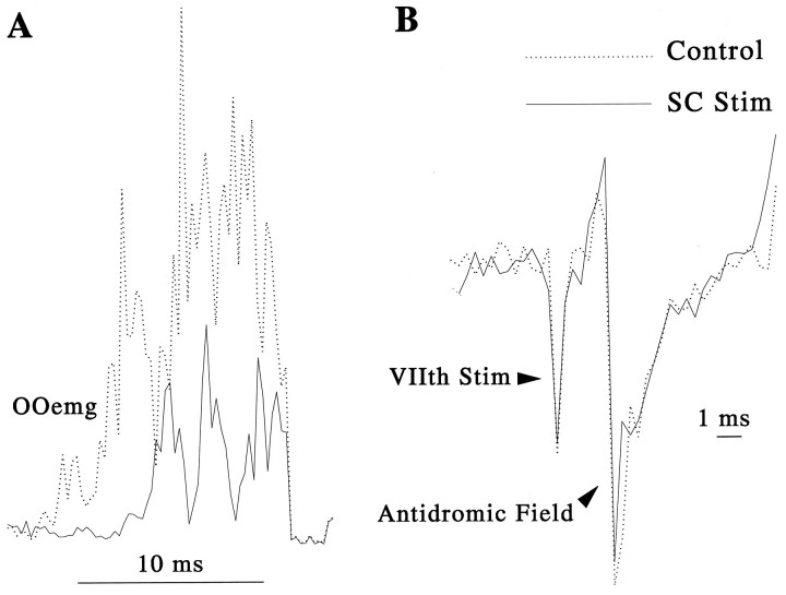
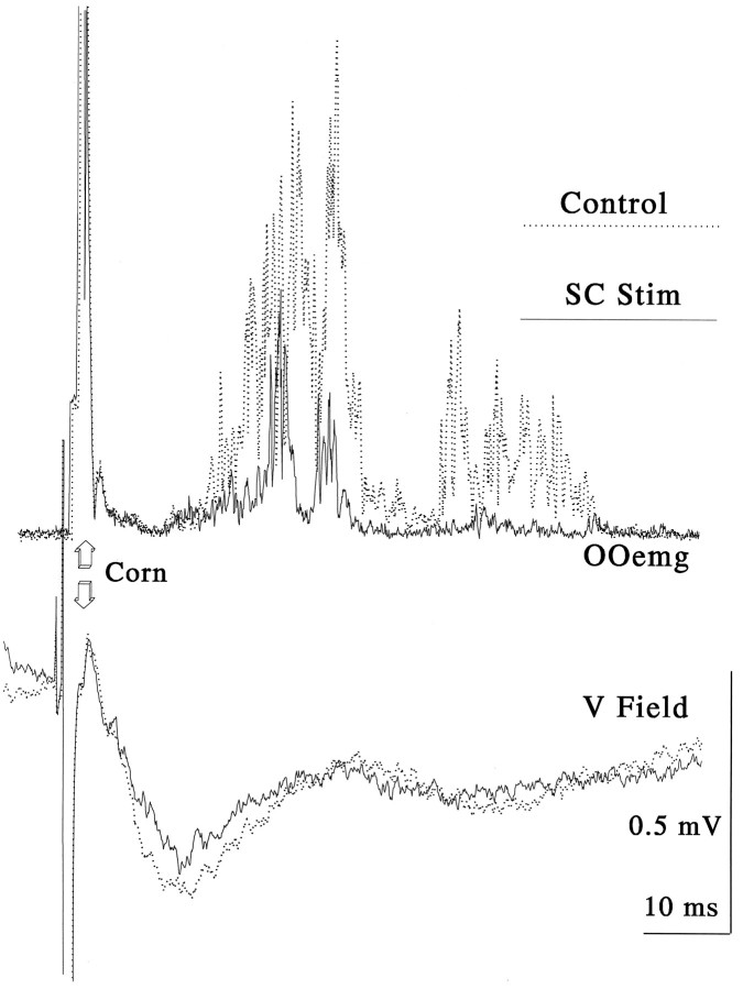
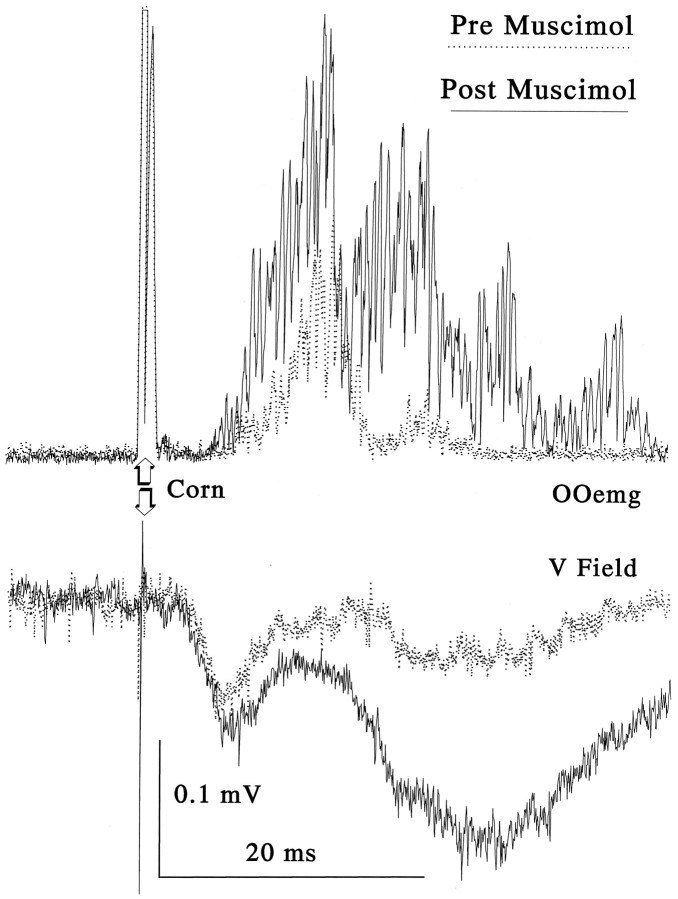
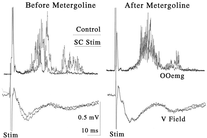
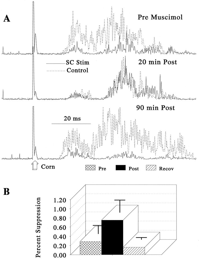
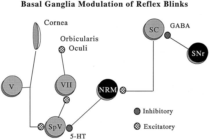
Hyperexcitable reflex blinks are a cardinal sign of Parkinson's disease. The first step in the circuit linking the basal ganglia and brainstem reflex blink circuits is the inhibitory nigrostriatal pathway (Basso et al., 1996). The current study reports the circuits linking the superior colliculus (SC) to trigeminal reflex blink circuits. Microstimulation of the deep layers of the SC suppresses subsequent reflex blinks at a latency of 5.4 msec. This microstimulation does not activate periaqueductal gray antinociceptive circuits. The brainstem structure linking SC to reflex blink circuits must suppress reflex blinks at a shorter latency than the SC and produce the same effect on reflex blink circuits as SC stimulation, and removal of the structure must block SC modulation of reflex blinks. Only the nucleus raphe magnus (NRM) meets these requirements. NRM microstimulation suppresses reflex blinks with a latency of 4.4 msec. Like SC stimulation, NRM microstimulation reduces the responsiveness of the spinal trigeminal nucleus. Finally, blocking the receptors for the NRM transmitter serotonin eliminates SC modulation of reflex blinks, and muscimol inactivation of the NRM transiently prevents SC modulation of reflex blinks. Thus, the circuit through which the basal ganglia modulates reflex blinking is (1) the substantia nigra pars reticulata inhibits SC neurons, (2) the SC excites tonically active NRM neurons, and (3) NRM neurons inhibit spinal trigeminal neurons involved in reflex blink circuits.


Figures

Fig. 1.
Fig. 1.
Effect of the delay between the onset of the SC stimulation and the occurrence of the corneal stimulus on reflex blink magnitude. A, OOemg response to a corneal stimulus (Corn) with (bottom trace) and without (top trace) a preceding 40 μA, 70 msec, 200 Hz train of SC stimulation (black bar) that terminated 5 msec before the corneal stimulus. Each trace is the average of five rectified blinks.B, The diagram illustrates stimulus conditions. The corneal stimulus (Corn) occurred 100 msec (−100 msec), 80 msec (−80 msec), 70 msec (−70 msec) after the onset of a 70 msec train of SC stimulation (SC Stim) or 10 msec (10 msec)before the onset of SC stimulation. C, Effect of SC stimulation on reflex blink magnitude as a function of time between corneal stimulation and the onset of a 70 msec train of SC stimulation (▾) compared to trials without superior colliculus stimulation (•). All data are normalized to the mean magnitude of all blinks at all delays without SC stimulation. Each point is the mean of 25 blinks (5 blinks from 5 animals), and the error bars are SEM.
Fig. 2.
Fig. 2.
Suppression of reflex blinks by NRM microstimulation. A, Orbicularis oculi response to a corneal stimulus (Corn) with (solid line) and without (dotted line) a preceding 70 msec, 200 Hz, 15 μA stimulus train to the nucleus raphe magnus (Raphe Stim) that ended 5 msec before the corneal stimulus. Each trace is the average of five rectified responses. B, Group data from four animals (at least 5 blinks per condition per animal) illustrating OOemg magnitude with preceding NRM microstimulation (solid bar, Stim) relative to OOemg blink magnitude without NRM stimulation (hatched bar, Con). Error bars are SEM.
Fig. 3.
Fig. 3.
A single stimulus to the superior colliculus or the nucleus raphe magnus transiently decreases reflex blink amplitude.A, Unfiltered OOemg response to corneal stimulation with (solid line, SC Stim) and without (dotted line, Unstim) a single 80 μsec pulse to the superior colliculus (SC Stim, dashed vertical line). Traces are unfiltered (top traces) or filtered (bottom traces) at 250 Hz to facilitate comparison of the stimulated and unstimulated records. Each trace is the average of five rectified OOemg responses.B, A single stimulus of the nucleus raphe magnus transiently decreases blink amplitude at a shorter latency than SC stimulation. OOemg response to corneal stimulation with (solid line, NRM Stim) and without (dotted line, Unstim) a single 80 μsec pulse to the nucleus raphe magnus (NRM Stim, dashed vertical line). Traces are unfiltered (top traces) or filtered (bottom traces) at 250 Hz to facilitate comparison of the stimulated and unstimulated records. Each trace is the average of five rectified OOemg responses.
Fig. 4.
Fig. 4.
Effect of trains of SC microstimulation on facial nucleus antidromic field potentials. A, OOemg response to corneal stimulation at the intensity used to assess OO motoneuron excitability with (solid line) and without (dashed line) preceding, 70 msec train of SC stimulation. B, Antidromic field potential from the OO subdivision of the facial nucleus evoked by stimulation of the zygomatic branch of the facial nerve with (solid line) and without (dashed line) preceding SC stimulation. Each trace is the average of 10 responses.
Fig. 5.
Fig. 5.
Effect of a train of SC stimulation on simultaneously recorded OOemg activity and trigeminal field potentials evoked by a corneal stimulus (⇓ Corn). Top traces are superimposed orbicularis oculi emg (OOemg) responses, and bottom traces are superimposed trigeminal field potentials (V Field). Solid linesshow trials with a preceding, 70 msec train of SC stimulation (SC Stim) that terminated 5 msec before the corneal stimulus.Dotted lines show records from unstimulated trails (Control). Each trace is an average of 10 responses.
Fig. 6.
Fig. 6.
Effect of a microinjection of muscimol into the SC stimulation on simultaneously recorded OOemg activity and trigeminal field potentials evoked by a corneal stimulus (⇑ Corn).Top traces are superimposed orbicularis oculi emg (OOemg) responses, and bottom traces are superimposed trigeminal field potentials (V Field).Solid lines show trials after a 0.5 μl injection of 1.0% muscimol into the contralateral SC (Post Muscimol).Dotted lines show records collected before the muscimol injection (Pre Muscimol). Each trace is an average of 12 responses.
Fig. 7.
Fig. 7.
Systemic metergoline injections block SC suppression of orbicularis oculi (OOemg) and trigeminal field potentials (V Field) evoked by corneal stimulation (Stim). Simultaneously recorded OOemg (top traces) and trigeminal field responses (bottom traces) to a corneal stimulus with (solid line, SC Stim) and without (dotted line, Control) a preceding, 70 msec SC stimulation that terminated 5 msec before the corneal stimulus before (left records) and after (right records) a 6 mg/kg injection of metergoline. Each trace is an average of 10 rectified responses.
Fig. 8.
Fig. 8.
Schematic coronal hemisections through the trigeminal nucleus of the rat brain (Swanson, 1992) illustrating the site of trigeminal field potential recordings. Values indicate distance (in mm) from bregma. Black dots show the location of trigeminal field potential recording sites. Black squaresare sites where data for Figures 5 and 6 were collected. IO, Inferior olive; LRN, lateral reticular nucleus;spVc, spinal trigeminal nucleus caudalis subdivision;spVi, spinal trigeminal nucleus interpolaris subdivision;XII, hypoglossal nucleus.
Fig. 9.
Fig. 9.
Effect of microinjections of muscimol into the nucleus raphe magnus on suppression of corneal evoked blinks by SC stimulation. A, OOemg responses to corneal stimulation with (dashed lines) and without (solid lines) a preceding, 70 msec train of SC stimulation that terminated 5 msec before a corneal stimulus (⇑ Corn) before (top trace, Pre Muscimol), 20 min after (middle traces, 20 min Post), and 90 min after (bottom traces, 90 min Post) a 50 nl injection of 1.0% muscimol into the NRM. Each trace is the mean of 10 responses. B, Group data showing the magnitude of blink reflex suppression with SC stimulation before (hatched bar, Pre), after (black bar, Post), and after recovery (striped bar, Recov) from a microinjection of muscimol into the NRM. In the pre and post condition, the bars show the averages of at least 60 blinks, 10 from each of 6 animals. In the recovery condition, the bars show averages of at least 20 blinks, 10 from each of two of these 6 animals. Error bars are SEM.
Fig. 10.
Fig. 10.
Location of muscimol injections in the nucleus raphe magnus experiments. Black squares show injection sites that reduced SC suppression of trigeminal reflex blinks within 10 min.Filled circles show injection sites where reduction of SC blink suppression occurred between 30 and 45 min after the injection. The open circles identify ineffective injection sites.gVII, Genu of the facial nerve; NPc, nucleus pontis caudalis; NRM, nucleus raphe magnus.
Fig. 11.
Fig. 11.
Circuit linking the basal ganglia and the trigeminal reflex blink circuit. NRM, Nucleus raphe magnus;SC, superior colliculus; SNr, substantia nigra pars reticulata; SpV, spinal trigeminal nucleus;V, trigeminal ganglion; VII, facial motoneurons.

References

    1. Auerbach S, Fornal C, Jacobs B. Response of serotonin containing neurons in nucleus raphe magnus to morphine, noxious stimuli, and periaqueductal gray stimulation in freely moving cats. Exp Neurol. 1985;88:609–628. - PubMed
    1. Basso MA, Strecker RE, Evinger C. Midbrain 6-hydroxdopamine lesions modulate blink reflex excitability. Exp Brain Res. 1993;94:88–96. - PubMed
    1. Basso MA, Powers AS, Evinger C. An explanation for blink reflex hyperexcitability in Parkinson’s disease. I. Superior colliculus. J Neurosci. 1996;16:7308–7317. - PMC - PubMed
    1. Bronstein AM, Kennard C. Predictive ocular motor control in Parkinson’s disease. Brain. 1985;108:925–940. - PubMed
    1. Browne PA, Goldberg LJ. Direct evidence of presynaptic inhibition of a trigeminal reflex. Brain Res. 1978;143:369–372. - PubMed

Publication types

LinkOut - more resources