

Magnetic resonance imaging study of hippocampal volume in chronic, combat-related posttraumatic stress disorder
- PMID: 8931911
- PMCID: PMC2910907
- DOI: 10.1016/S0006-3223(96)00229-6
Magnetic resonance imaging study of hippocampal volume in chronic, combat-related posttraumatic stress disorder
Abstract
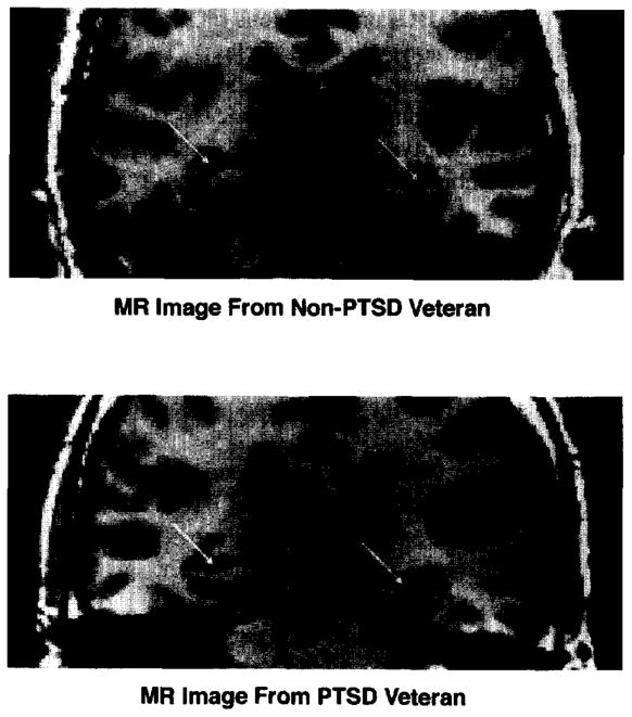
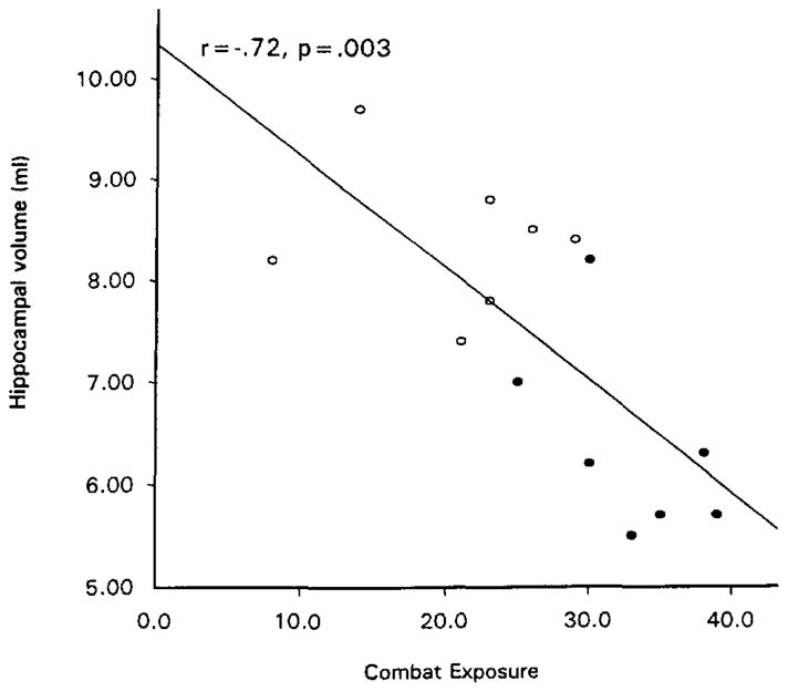
This study used quantitative volumetric magnetic resonance imaging techniques to explore the neuroanatomic correlates of chronic, combat-related posttraumatic stress disorder (PTSD) in seven Vietnam veterans with PTSD compared with seven nonPTSD combat veterans and eight normal nonveterans. Both left and right hippocampi were significantly smaller in the PTSD subjects compared to the Combat Control and Normal subjects, even after adjusting for age, whole brain volume, and lifetime alcohol consumption. There were no statistically significant group differences in intracranial cavity, whole brain, ventricles, ventricle:brain ratio, or amygdala. Subarachnoidal cerebrospinal fluid was increased in both veteran groups. Our finding of decreased hippocampal volume in PTSD subjects is consistent with results of other investigations which utilized only trauma-unexposed control groups. Hippocampal volume was directly correlated with combat exposure, which suggests that traumatic stress may damage the hippocampus. Alternatively, smaller hippocampi volume may be a pre-existing risk factor for combat exposure and/or the development of PTSD upon combat exposure.
Figures
References
-
- Axelson DA, Doraiswamy PM, McDonald WM, Boyko OB, Tupler LA, Patterson LJ, Nemeroff CB, Ellinwood EH, Krishna KRR. Hypercortisolemia and hippocampal changes in depression. Psychiatry Res. 1993;47:163–173. - PubMed
-
- Baydena L, Branchey MH, Noumair D. Age of alcoholism onset I: Relationship to psychopathology. Arch Gen Psychiatry. 1989;46:225–230. - PubMed
-
- Benton AB. Benton Visual Retention Test. 5. San Antonio: Harcourt Brace Jovanovich, Inc. (The Psychological Corporation); 1992.
-
- Blake DD, Weathers FW, Nagy LM, Kaloupek DG, Gusman FD, Charney DS, Keane TM. The development of a clinician-administered PTSD scale. J Traumatic Stress. 1995;8:75–90. - PubMed
-
- Bogerts B, Lieberman JA, Ashtari M, Bilder RM, Degreef G, Lerner G, Johns C, Masiar S. Hippocampus-amygdala volumes and psychopathology in chronic schizophrenia. Biol Psychiatry. 1993;33:236–246. - PubMed
Publication types
MeSH terms
Grants and funding
LinkOut - more resources
Full Text Sources
Other Literature Sources
Medical
