

Reproducibility of nerve fiber layer thickness measurements using optical coherence tomography
- PMID: 8942887
- PMCID: PMC1939724
- DOI: 10.1016/s0161-6420(96)30410-7
Reproducibility of nerve fiber layer thickness measurements using optical coherence tomography
Abstract
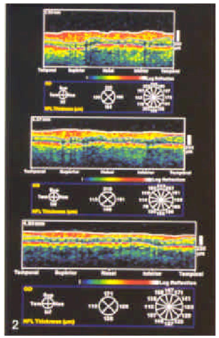
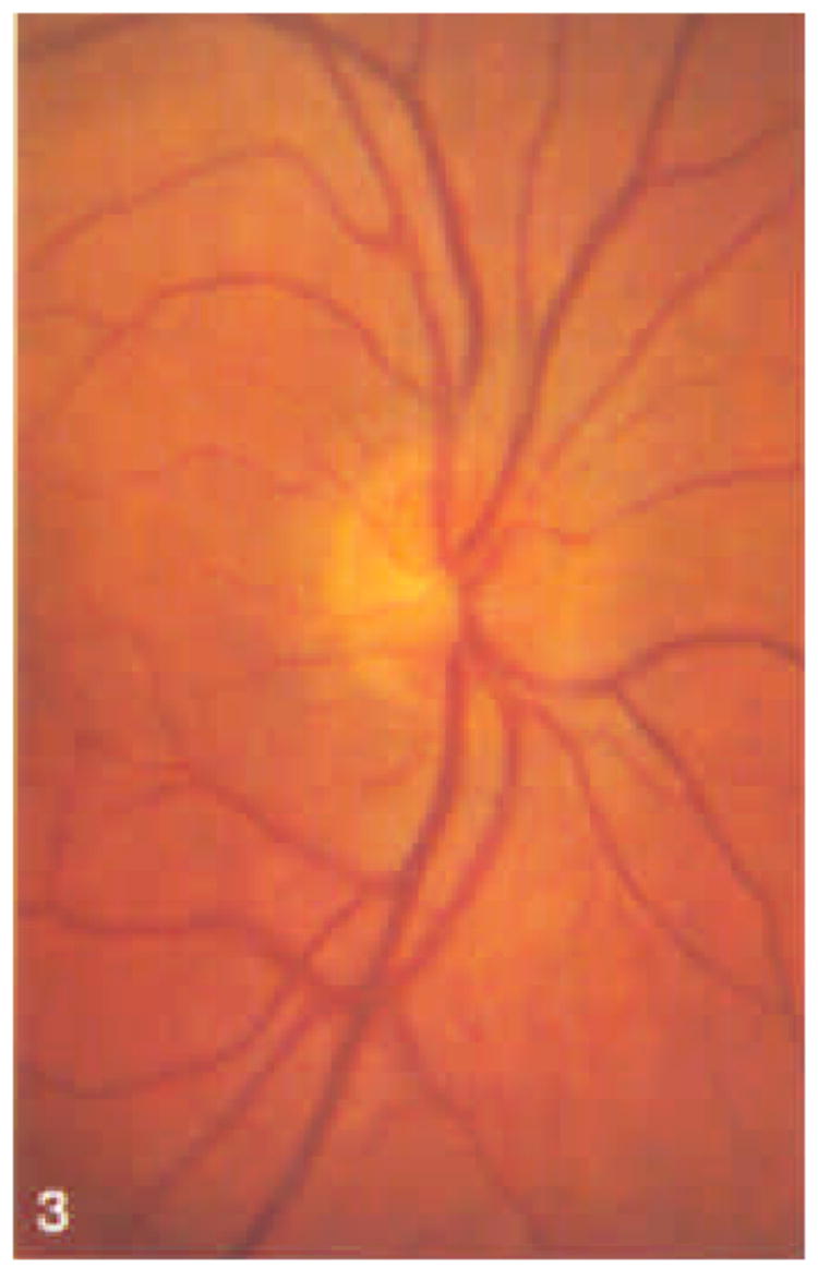
Purpose: Optical coherence tomography (OCT) is a new technology that uses near-infrared light in an interferometer to produce approximately 10-microns resolution cross-sectional images of the tissue of interest. The authors performed repeated quantitative assessment of nerve fiber layer thickness in individuals with normal and glaucomatous eyes, and they evaluated the reproducibility of these measurements.
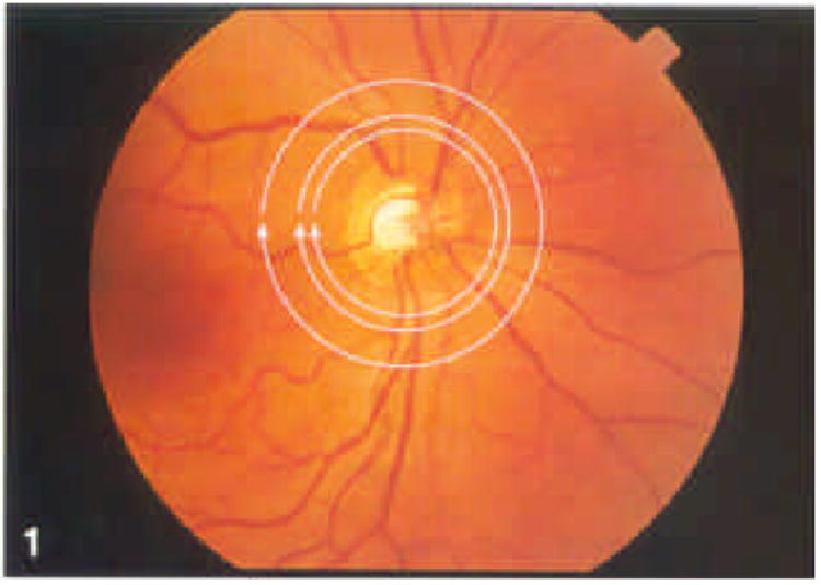
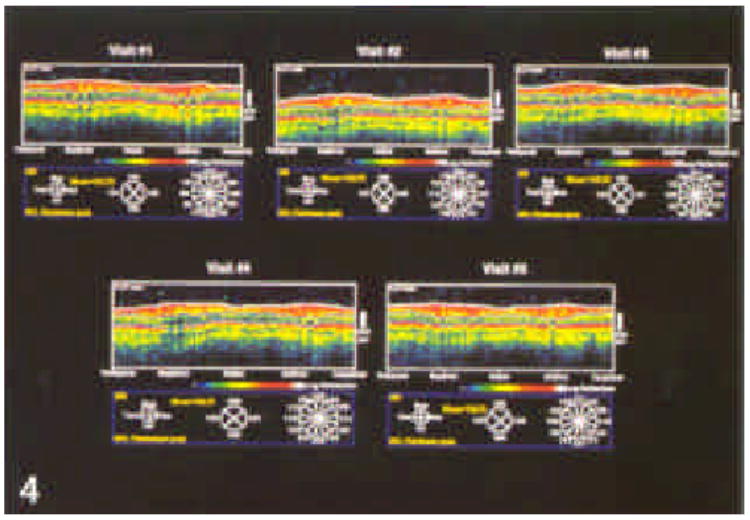
Methods: The authors studied 21 eyes of 21 subjects by OCT. Each subject underwent five repetitions of a series of scans on five separate occasions within a 1-month period. Each series consisted of three circular scans around the optic nerve head (diameters, 2.9, 3.4, and 4.5 mm). Each series was performed separately using internal (fixation with same eye being studied) and external (fixation with contralateral eye) fixation techniques. The eye studied and the sequence of testing were assigned randomly.
Results: Internal fixation (IF), in general, provides a slightly higher degree of reproducibility than external fixation (EF). Reproducibility was better in a given eye on a given visit than from visit to visit. Reproducibility as measured by intraclass correlation coefficients were as follows: circle diameter (CD), 2.9 mm, 0.51/0.57 (normal/glaucoma) (IF), 0.43/0.54 (EF); CD, 3.4 mm, 0.56/0.52 (IF), 0.43/0.61 (EF); CD, 4.5 mm, 0.53/0.43 (IF), 0.42/0.49 (EF).
Conclusions: Nerve fiber layer thickness can be reproducibly measured using OCT. Internal is superior to external fixation; each circle diameter tested provides adequate reproducibility.
Figures

Comment in
-
Reproducibility of nerve fiber layer thickness measurements.Ophthalmology. 1997 Oct;104(10):1530-1. doi: 10.1016/s0161-6420(97)30100-6. Ophthalmology. 1997. PMID: 9331186 No abstract available.
References
-
- Izatt JA, Hee MR, Huang D, et al. Ophthalmic diagnostics using optical coherence tomography. In: Ren Q, Pavel JM, editors. Proceedings of Ophthalmic Technologies III; January 1992; Los Angeles, CA. Bellingham, WA: SPIE; 1993. pp. 136–44. (Progress in biomedical optics; Proceedings of SPIE; vol. 1877.)
-
- Swanson EA, Izatt JA, Hee MR, et al. In vivo retinal imaging using optical coherence tomography. Opt Lett. 1993;18:1864–6. - PubMed
-
- Hee MR, Izatt JA, Swanson EA, et al. Optical coherence tomography of the human retina. Arch Ophthalmol. 1995;113:325–32. - PubMed
-
- Izatt JA, Hee MR, Swanson EA, et al. Micrometer-scale resolution imaging of the anterior eye in vivo with optical coherence tomography. Arch Ophthalmol. 1994;112:1584–9. - PubMed
Publication types
MeSH terms
Grants and funding
LinkOut - more resources
Full Text Sources
Other Literature Sources
Medical
