Review
. 1996 Nov 26;93(24):13500-7.
doi: 10.1073/pnas.93.24.13500.

Functional organization of the hippocampal memory system

Affiliations
Review

Functional organization of the hippocampal memory system

H Eichenbaum et al. Proc Natl Acad Sci U S A. .

Abstract

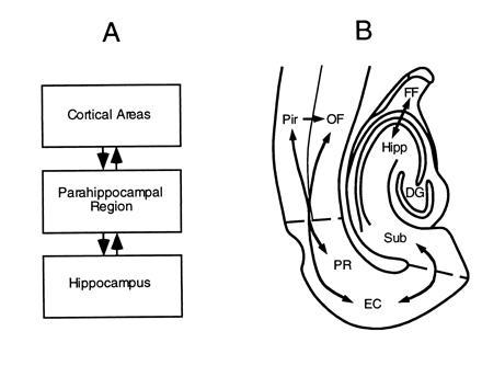
In humans declarative or explicit memory is supported by the hippocampus and related structures of the medial temporal lobe working in concert with the cerebral cortex. This paper reviews our progress in developing an animal model for studies of cortical-hippocampal interactions in memory processing. Our findings support the view that the cortex maintains various forms of memory representation and that hippocampal structures extend the persistence and mediate the organization of these codings. Specifically, the parahippocampal region, through direct and reciprocal interconnections with the cortex, is sufficient to support the convergence and extended persistence of cortical codings. The hippocampus itself is critical to the organization cortical representations in terms of relationships among items in memory and in the flexible memory expression that is the hallmark of declarative memory.


Figures

Figure 1
Figure 1
(A) Simple schematic diagram of cortical–hippocampal connections. (B) Outline of a horizontal rat brain section illustrating the locations and flow of information between components of the hippocampus, parahippocampal region, and adjacent cortical areas. DG, dentate gyrus; EC, entorhinal cortex; FF, fimbria–fornix; Hipp, hippocampus proper; OF, orbitofrontal cortex; Pir, perirhinal cortex; PR, perirhinal cortex; Sub, subiculum.
Figure 2
Figure 2
Odor responses of olfactory cortex neurons. The dark bars represent neural responses to S+ odors and striped bars represent responses to S− odors. OF, orbitofrontal.
Figure 3
Figure 3
Short-term memory correlates of the activity of olfactory cortical neurons. Each panel illustrates the neural response of a cell to various current odors depending upon the stimulus on the preceding trial. OF, orbitofrontal.
Figure 4
Figure 4
Neural correlates of odor–odor associations in the olfactory cortex. See text for description.
Figure 5
Figure 5
Neural activity in the parahippocampal region associated with DNMS performance. (Left) Responses of the cell during the 2 sec following the onset of different odors. (Right) Activity during the last 2 sec of the that delay period—i.e., prior to the onset of the subsequent odor cue. Note greater activity for odor 5 in both periods.
Figure 6
Figure 6
Neural activity in the hippocampus associated with DNMS performance. See text for description.
Figure 7
Figure 7
(A) Training and testing on a “naturalistic” odor–odor association. In phase I a “demonstrator” rat is given a distinctively scented food. Then, in phase II, the demonstrator is presented to an experimental subject for a brief period of social interaction. During this experience subjects associate two odors carried on the demonstrator’s breath, the distinctive food odor and carbon disulfide (CS2), a natural constituent of rats’ breath (ref. ; phase II). In phase III, to test memory for the food odor–CS2 association, subjects are presented with the same food or another distinctively scented food, either immediately or after a 24-hr delay. After this training normal rats show a strong selection preference for the trained food odor. (B) The effects of hippocampal lesions on performance in the retention test. Retention was intact immediately after learning but severely impaired during a 24-hr retention test.
Figure 8
Figure 8
Inferential expression of odor–odor associations. (A) Schematic diagram of paired associate training and probe testing. Letters represent odor stimulus items; arrows without question marks indicate trained pairings, whereas arrows with question marks indicate expected transitive and symmetrical choices. Rats are first trained on two overlapping sets of paired associates (Left). Then (Right) they are tested for inferential expression in two ways. In the test for transitivity, they are presented with one of two sample cues from the first training set and the required to select between the choice cues from the second set, based on the shared associates of these items. In the test for symmetry or “reversibility” of the associations, they are presented with one of two choice cues from the second set and required to select the appropriate sample cue from that set. (B) Errors to criterion on acquisition of the two sets of paired associates for sham-operated and hippocampal subjects. (C) Preference indices on the test for transitive inference. For these probe trials a preference score was calculated as (XY)/(X + Y), where X and Y were the digging times in the transitive and alternate choices, respectively. (D) Preference indices on the test for symmetrical expression.

References

    1. Gray J A. The Neuropsychology of Anxiety: An Enquiry into the Functions of the Septo-Hippocampal System. New York: Oxford Univ. Press; 1982.
    1. Halgren E. In: Neuropsychology of Memory. Butters N, Squire L R, editors. New York: Guilford; 1984. pp. 165–182.
    1. Rolls E. In: Neural Models of Plasticity: Theoretical and Empirical Approaches. Byrne J H, Berry W O, editors. New York: Academic; 1989. pp. 240–265.
    1. Squire L R, Cohen N J, Nadel L. In: Memory Consolidation. Weingartner H, Parker E, editors. Hillsdale, NJ: Erlbaum; 1984. pp. 185–210.
    1. Teyler T J, DiScenna P. Behav Neurosci. 1986;100:147–154. - PubMed

Publication types