Review
. 1996 Nov 26;93(24):13508-14.
doi: 10.1073/pnas.93.24.13508.

Involvement of the amygdala in memory storage: interaction with other brain systems

Affiliations
Review

Involvement of the amygdala in memory storage: interaction with other brain systems

J L McGaugh et al. Proc Natl Acad Sci U S A. .

Abstract

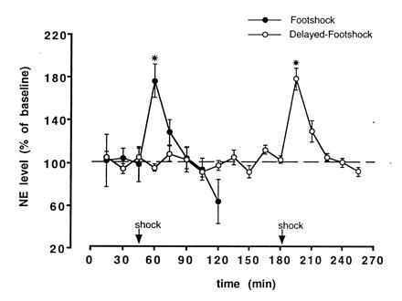
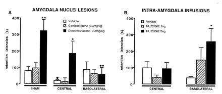
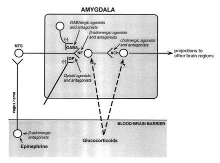
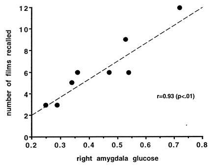
There is extensive evidence that the amygdala is involved in affectively influenced memory. The central hypothesis guiding the research reviewed in this paper is that emotional arousal activates the amygdala and that such activation results in the modulation of memory storage occurring in other brain regions. Several lines of evidence support this view. First, the effects of stress-related hormones (epinephrine and glucocorticoids) are mediated by influences involving the amygdala. In rats, lesions of the amygdala and the stria terminalis block the effects of posttraining administration of epinephrine and glucocorticoids on memory. Furthermore, memory is enhanced by posttraining intraamygdala infusions of drugs that activate beta-adrenergic and glucocorticoid receptors. Additionally, infusion of beta-adrenergic blockers into the amygdala blocks the memory-modulating effects of epinephrine and glucocorticoids, as well as those of drugs affecting opiate and GABAergic systems. Second, an intact amygdala is not required for expression of retention. Inactivation of the amygdala prior to retention testing (by posttraining lesions or drug infusions) does not block retention performance. Third, findings of studies using human subjects are consistent with those of animal experiments. beta-Blockers and amygdala lesions attenuate the effects of emotional arousal on memory. Additionally, 3-week recall of emotional material is highly correlated with positronemission tomography activation (cerebral glucose metabolism) of the right amygdala during encoding. These findings provide strong evidence supporting the hypothesis that the amygdala is involved in modulating long-term memory storage.


Figures

Figure 1
Figure 1
Freely moving rats with a microdialysis probe inserted into a cannula implanted in the amygdala (tip aimed at the border between the Central and Basolateral nuclei) received a single footshock (0.55 mA, 1.0 s) either 45.5 min (n = 5) or 180.5 min (n = 4) after being placed in an apparatus with a grid floor. Dialyzate samples were collected every 15 min and immediately injected into an HPLC with coulometric detection optimized for detection of NE. NE concentration is represented as mean (±SEM) of basal level prior to footshock. NE concentrations in the dialyzate increased to ≈75% above baseline (∗, P < 0.001) following the footshock and returned to baseline within 30 min. [Reproduced with permission from ref. (Copyright 1996, Harcourt Brace.]
Figure 2
Figure 2
Step-through latencies (mean ± SEM) for a 48-h inhibitory avoidance retention test. (A) Rats with sham, or lesions of either the central or basolateral nucleus had been treated with corticosterone, dexamethasone, or vehicle immediately following training. (B) Rats received posttraining microinfusions of the glucocorticoid agonist RU 28362 into the central or basolateral nucleus. ∗, P < 0.05; ∗∗, P < 0.01 as compared with the corresponding vehicle group; •, P < 0.05 as compared with the corresponding sham lesion-vehicle group; ♦♦, P < 0.01 as compared with the corresponding sham lesion-dexamethasone group. [Reproduced with permission from ref. (Copyright 1996, Harcourt Brace.]
Figure 3
Figure 3
Schematic summarizing the interactions of neuromodulatory systems influencing memory storage suggested by the findings of our experiments. Epinephrine acts at peripheral β-adrenergic receptors located on vagal afferents projecting to the NTS. Activation of the NTS induces NE release in the amygdala. The peripherally acting β-adrenergic antagonist sotalol blocks epinephrine effects. Centrally acting noradrenergic agonists (clenbuterol and dipivefrin) directly activate NE receptors in the amygdala. Opiate and GABAergic agonists inhibit NE release and opiate and GABAergic antagonists induce NE release by blocking the inhibition. Centrally acting β-adrenergic antagonists (e.g., propranolol) block the activation of NE receptors in the amygdala and, thus, block all neuromodulatory influences affecting NE release. A subsequent step involves activation of muscarinic cholinergic receptors within the amygdala. Drugs affecting muscarinic cholinergic activation block the effects of drugs influencing noradrenergic activation. Glucocorticoids act at several sites. The modulatory effects of hormones and drugs on memory storage are mediated by influences on other brain systems.
Figure 4
Figure 4
Activity of the amygdala related to emotional memory in healthy human subjects. Scatterplot shows the relationship between relative glucose metabolic rate of the right amygdala in subjects viewing a series of emotionally arousing (aversive) film clips and long-term memory of those clips. Relative glucose metabolic rate in the right amygdala was highly correlated (P < 0.01) with the number of films recalled. [Reproduced with permission from Cahill et al. (22) (Copyright 1996, National Acadeny of Sciences).]

References

    1. Weiskrantz L. J Comp Physiol Psychol. 1956;49:381–391. - PubMed
    1. Aggleton J P, editor. The Amygdala. New York: Wiley–Liss; 1992.
    1. Scoville W B, Milner B. J Neurol Neurosurg Psychiatry. 1957;20:11–21. - PMC - PubMed
    1. Zola-Morgan S, Squire L R, Amaral D G. J Neurosci. 1989;9:1922–1936. - PMC - PubMed
    1. Davis M. Annu Rev Neurosci. 1992;15:353–375. - PubMed

Publication types