. 1996 Nov 26;93(24):13571-6.
doi: 10.1073/pnas.93.24.13571.

YY1 transcriptional initiator: protein interactions and association with a DNA site containing unpaired strands

Affiliations

YY1 transcriptional initiator: protein interactions and association with a DNA site containing unpaired strands

A Usheva et al. Proc Natl Acad Sci U S A. .

Abstract

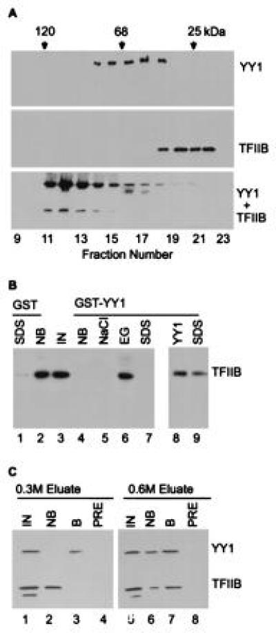
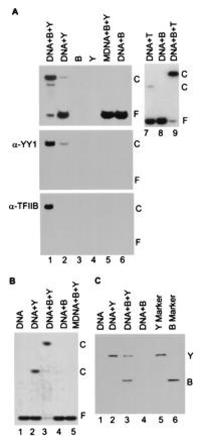
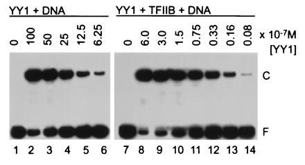
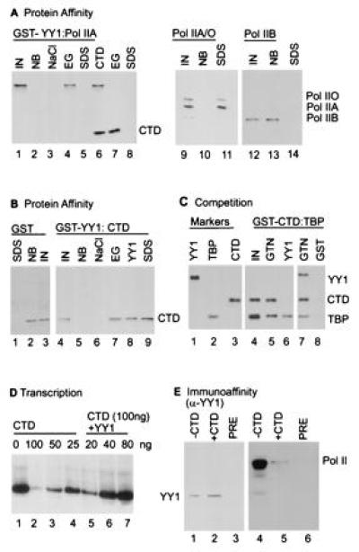
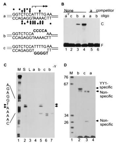
The Ying-Yang 1 protein (YY1) DNA-binding site functions as an initiator element at which YY1, transcription factor IIB (TFIIB), and RNA polymerase II sponsor basal transcription from a supercoiled DNA template. We show that TFIIB binds to YY1, stabilizing its interaction with DNA, and YY1 contacts the large subunit of polymerase II, directing it to the initiation site. YY1 directs initiation from linear DNA containing mismatched sequences within its binding site, leading us to infer that supercoiling facilitates the separation of DNA strands and to suggest that YY1 likely remains bound to the start site as DNA strands separate during initiation. These results provide a mechanistic basis for transcriptional initiation directed by YY1 in the absence of the TATA box-binding protein.


Figures

Figure 1
Figure 1
TFIIB binds to YY1. (A) TFIIB binds to YY1 in solution. Purified TFIIB and YY1 were subjected to gel filtration chromatography alone or after mixing, and localized in the elution profile by protein blot assay using polyclonal antibody (18) to YY1 (Top), TFIIB (Middle), or a mixture of the antibodies (Bottom). Chymotrypsinogen A (25 kDa), BSA (68 kDa), and β-galactosidase (120 kDa) were markers. (B) TFIIB binds to GST–YY1. TFIIB was monitored by protein blot assay. Lanes: IN, TFIIB input to each reaction; NB, TFIIB not bound; NaCl, TFIIB eluted in buffer containing 1.0 M NaCl; EG, TFIIB eluted in buffer containing 50% ethylene glycol plus 100 mM NaCl; SDS, TFIIB eluted by boiling in buffer with detergent, either immediately after application to the affinity matrix (lanes 1 and 9) or following elution with ethylene glycol (lane 7); YY1, TFIIB eluted in buffer containing 60 μg/ml YY1. (C) Copurification of TFIIB and YY1 from a HeLa cell nuclear extract. Two fractions from a single-stranded DNA cellulose column (0.3 M and 0.6 M NaCl) containing both YY1 and TFIIB were assayed on a YY1-specific IgG matrix. TFIIB and YY1 were detected by protein blot assay. Lanes: IN, TFIIB and YY1 input to each reaction; NB, input which is not bound; B, input which is bound; PRE, input bound by preimmune IgG. Figures were produced using photoshop and freehand software.
Figure 2
Figure 2
TFIIB forms a complex on DNA with YY1. (A) Band-shift assay of YY1–DNA interactions in a polyacrylamide gel. The components added to band-shift assays are indicated at the top of the autoradiograms. Lanes: B, TFIIB; Y, YY1; T, TBP; DNA, a 22-bp oligonucleotide with the YY1 binding site present at +1 in the AAV P5 promoter or a 20-bp oligonucleotide with the TATA motif present in the adenovirus major late promoter; and MDNA, a 22-bp derivative of the YY1-binding site to which YY1 cannot bind (14). (Top) Band-shift assay displays an autoradiogram where 32P-labeled probe DNA is detected. (Middle and Bottom) Protein blots using antibody to YY1 (α-YY1) or TFIIB (α-TFIIB). Reactions for band-shift assays in which proteins were transferred to membranes received 25-fold more of each protein than was used in standard band-shift assays. The positions of free DNA, F, and protein–DNA complexes, C, are indicated. (B) Band-shift assay in an agarose gel showing that TFIIB, YY1 and DNA form a complex. Components added to assays are as in A. The YY1–TFIIB–DNA complex was unstable during analysis in agarose, as evidenced by the substantial reduction in the amount of free DNA without a concomitant increase in the band corresponding to the complex (lane 3). (C) Analysis of proteins present in shifted complexes. Complexes present in the agarose gel displayed in B were excised, and their protein constituents were analyzed by electrophoresis in an SDS-containing polyacrylamide gel. Purified YY1 (Y) and TFIIB (B) were included as markers.
Figure 3
Figure 3
TFIIB increases the apparent affinity of YY1 for its DNA-binding site. The amount of YY1 added to each band-shift reaction is recorded above the lanes, and the positions of free DNA (F) and protein–DNA complexes (C) are indicated.
Figure 4
Figure 4
RNA pol II interacts with YY1. (A) The large subunit of RNA pol II binds to GST–YY1. RNA pol IIA (pol IIA) mixed RNA pol IIA and pol IIO (pol IIA/O) and RNA pol IIB (pol IIB) were tested for interaction with GST–YY1. Polymerase was monitored by protein blot assay using antibodies specific for the CTD (lanes 1–8) or the amino acid sequence encoded by exon 5 of the mRNA encoding the large subunit (lanes 9–14). Lanes: IN, polymerase input; NB, polymerase not bound; NaCl, polymerase eluted in buffer containing 1.0 M NaCl; EG, polymerase eluted in buffer containing 50% ethylene glycol plus 100 mM NaCl; CTD, polymerase eluted by inclusion of a CTD polypeptide (100 μg/ml) in the washing buffer; SDS, polymerase eluted by boiling in buffer with detergent, after elution with ethylene glycol (lanes 5 and 8) or immediately after loading onto GST–YY1 matrix (lanes 11 and 14). (B) The CTD binds to GST–YY1. The CTD polypeptide was detected by protein blot, and the designations for lanes are as for A. (C) YY1 competes with TBP for interaction with the CTD. TBP was bound to GST–CTD fusion protein on beads, complexes on beads were washed with binding buffer, TBP was displaced from the GST–CTD–TBP complex with binding buffer containing soluble YY1 (100 μl, 4 mg/ml), and reactants were assayed by protein blot using a mixture of antibodies to YY1, TBP, and CTD (lanes 1–3, 7, and 8) or TPB and CTD (lanes 4–6). YY1-specific antibody was excluded from the membrane including lane 6 because it contained a huge quantity of YY1. Lanes: IN, CTD–TBP complex input; GTN, elution with glutathionine; YY1, elution with YY1; GST, elution with GST protein without a fusion partner. (D) CTD inhibits transcription directed by the AAV P5 promoter in the presence of YY1, TFIIB, and pol IIA. Reactions received the indicated amounts of CTD polypeptide and GST–YY1 fusion protein. The 79-nt primer extension products are labeled. (E) Copurification of pol II and YY1 from a HeLa nuclear extract. Partially purified polymerase was incubated with YY1-specific antibody on beads in the presence or absence of excess CTD polypeptide, ±CTD. Material bound to YY1-specific antibody, α-YY1, was eluted and assayed by protein blot using either an antibody to YY1 (lanes 1 and 2) or an antibody to the polymerase (lanes 3 and 4).
Figure 5
Figure 5
YY1-directed transcription from linear DNA containing a mismatched sequence at the initiation site. (A) Sequences of oligonucleotides with normal and altered YY1 binding sites. Interactions between YY1 and its recognition site (37) are indicated for oligonucleotide a. (B) Band-shift assay comparing the ability of YY1 to interact with its normal recognition site and modified sites containing mismatched sequences. 32P-labeled and unlabeled competitor oligonucleotides are identified at the top of the lanes. Lanes: a−Y, received oligonucleotide but no YY1; F, free DNA; and C, YY1–DNA complexes. (C) In vitro transcription assayed by primer extension. Transcription reactions received as template supercoiled (S) or linear (L) plasmid DNA with the AAV P5 promoter, the indicated oligonucleotide, or oligonucleotide b in a reaction lacking YY1 (b−Y). Markers (M) were generated by chemical sequencing using the A > C reaction of oligonucleotide a; initiation sites and bands corresponding to YY1-dependent initiation events are designated with arrows. (D) In vitro transcription monitored by run off assay. Reactions received oligonucleotide a, b, or c as template. The sizes of marker DNAs are indicated in bases; specific transcripts originating from the YY1-directed initiation sites and nonspecific transcripts form the ends of the bubbles are labeled.

References

    1. Weis L, Reinberg D. FASEB J. 1992;6:3300–3309. - PubMed
    1. Hernandez N. Genes Dev. 1993;7:1291–1308. - PubMed
    1. Gill G. Curr Biol. 1994;4:374–376. - PubMed
    1. Smale S T, Baltimore D. Cell. 1989;57:103–113. - PubMed
    1. Javahery R, Khachi A, Lo K, Zenzie-Gregory B, Smale S T. Mol Cell Biol. 1994;14:116–127. - PMC - PubMed

Publication types

LinkOut - more resources