. 1996 Nov 26;93(24):13629-34.
doi: 10.1073/pnas.93.24.13629.

hMSH2 forms specific mispair-binding complexes with hMSH3 and hMSH6

Affiliations

hMSH2 forms specific mispair-binding complexes with hMSH3 and hMSH6

S Acharya et al. Proc Natl Acad Sci U S A. .

Abstract

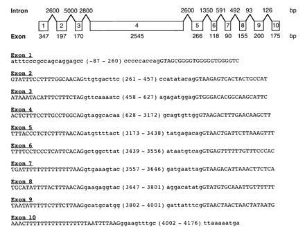
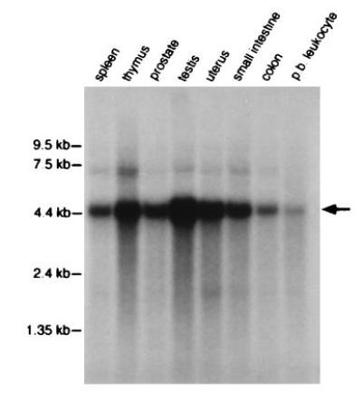
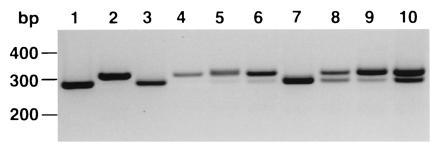
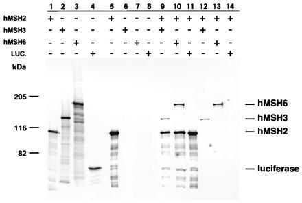
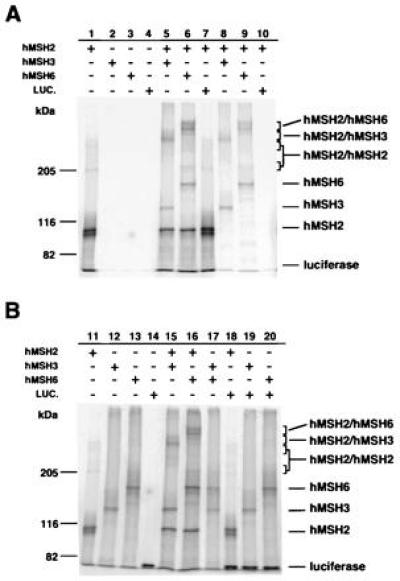
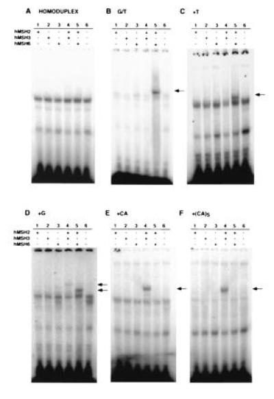
The genetic and biochemical properties of three human MutS homologues, hMSH2, hMSH3, and hMSH6, have been examined. The full-length hMSH6 cDNA and genomic locus were isolated and characterized, and it was demonstrated that the hMSH6 gene consisted of 10 exons and mapped to chromosome 2p15-16. The hMSH3 cDNA was in some cases found to contain a 27-bp deletion resulting in a loss of nine amino acids, depending on the individual from which the cDNA was isolated. hMSH2, hMSH3, and hMSH6 all showed similar tissue-specific expression patterns. hMSH2 protein formed a complex with both hMSH3 and hMSH6 proteins, similar to protein complexes demonstrated by studies of the Saccharomyces cerevisiae MSH2, MSH3, and MSH6. hMSH2 was also found to form a homomultimer complex, but neither hMSH3 nor hMSH6 appear to interact with themselves or each other. Analysis of the mismatched nucleotide-binding specificity of the hMSH2-hMSH3 and hMSH2-hMSH6 protein complexes showed that they have overlapping but not identical binding specificity. These results help to explain the distribution of mutations in different mismatch-repair genes seen in hereditary nonpolyposis colon cancer.


Figures

Figure 1
Figure 1
Organization of hMSH6 genomic locus and sequence of the intron region flanking each MSH6 exon. Boxes containing numbers 1 through 10 indicate the individual MSH6 exons and their relative sizes. The size of each exon is given below each exon and the size of each intron is given above the region between each pair of exons. The sizes of exons 1 and 10 are the sizes of the mRNA sequence upstream and downstream of the first and last introns, respectively; these sizes were calculated from the longest MSH6 cDNA sequence available. The first 20 nucleotides of each intron sequence up to the intron-exon junction is given in uppercase letters except for the 3′ side of the last intron, where additional sequence is given. The first 10 nucleotides of each exon sequence up to the intron-exon junction is given in lowercase letters. In addition, the sequence of the 5′ and 3′ ends of the cDNA, minus the poly(A) sequence, is also given in lowercase letters. The numbers in parentheses between intron sequences are the nucleotide coordinates of the exon sequences or cDNA sequences, assuming the A of the ATG is nucleotide 1. (Additional intron sequence including the complete sequence of some introns has been determined in all cases and is available on request from M.F.K. and R.K.)
Figure 2
Figure 2
Tissue expression of hMSH6 mRNA. A human multiple-tissue Northern blot (CLONTECH) was probed with the 32P-labeled complete cDNA clone of hMSH6 according to the manufacturer’s protocol. Colon refers to mucosal lining; p.b. leukocyte refers to peripheral blood leukocyte. The hMSH6 transcript is indicated by the arrow and corresponds to ≈4.5 kb. The amount of RNA loaded in each lane was adjusted to comparable levels as judged spectrophotometrically and by the levels of actin present (CLONTECH).
Figure 3
Figure 3
Analysis of hMSH3 27-bp deletion in various cDNAs. Tissue cDNAs from individuals or combined groups (CLONTECH) were used in a PCR reaction with primers flanking the region of the 27-bp deletion. The expected sizes of the products are 307 bp for the wild type and 280 bp for the deletion. Lane 1, MS3-12 HeLa hMSH3 cDNA clone; lane 2, HL60R hMSH3 cDNA clone; lane 3, HeLa cDNA; lane 4, liver cDNA from 40-year-old Caucasian male; lane 5, testis cDNA pooled from seven Caucasians aged 10–37; lane 6, placenta cDNA; lane 7, SW480 colorectal carcinoma cell line cDNA; lane 8, small intestine cDNA from a 22-year-old male; lane 9, bone marrow cDNA pooled from 24 Caucasian males and females aged 16–70; lane 10, kidney cDNA (unknown origin).
Figure 4
Figure 4
Immunoprecipitation of hMSH2, hMSH3, and hMSH6 protein complexes. In vitro transcribed and translated hMSH2, hMSH3, and hMSH6 were mixed and processed for immunoprecipitation (see Materials and Methods). Lanes 1–4, translations of hMSH2, hMSH3, hMSH6, and luciferase, respectively; lanes 5–8, immunoprecipitates of individual translated hMSH2, hMSH3, hMSH6, and luciferase, respectively; lane 9, co-immunoprecipitate of hMSH2 and hMSH3; lane 10, co-immunoprecipitate of hMSH2 and hMSH6; lane 11, co-immunoprecipitate of hMSH2 and luciferase; lane 12, co-immunoprecipitate of hMSH2 (unlabeled) and hMSH3; lane 13, co-immunoprecipitate of hMSH2 (unlabeled) and hMSH6; lane 14, co-immunoprecipitate of hMSH2 (unlabeled) and luciferase.
Figure 5
Figure 5
Chemical cross-link analysis of hMSH2, hMSH3, and hMSH6 protein complexes. The in vitro transcribed and translated proteins were mixed, cross-linked, and processed for immunoprecipitation (see Materials and Methods). (A) Lanes 1–4, immunoprecipitates of individually cross-linked hMSH2 (lane 1), hMSH3 (lane 2), hMSH6 (lane 3), and luciferase (lane 4); lanes 5–10, immunoprecipitates of the cross-linked complexes hMSH2 and hMSH3 (lane 5), hMSH2 and hMSH6 (lane 6), hMSH2 and luciferase (lane 7), hMSH2 (unlabeled) and hMSH3 (lane 8), hMSH2 (unlabeled) and hMSH6 (lane 9), hMSH2 (unlabeled) and luciferase (lane 10). (B) Lanes 11–18, cross-linked complexes before immunoprecipitation including hMSH2 (lane 11), hMSH3 (lane 12), hMSH6 (lane 13), luciferase (lane 14), hMSH2 and hMSH3 (lane 15), hMSH2 and hMSH6 (lane 16), hMSH3 and hMSH6 (lane 17), hMSH2 and luciferase (lane 18), hMSH3 and luciferase (lane 19), hMSH6 and luciferase (lane 20).
Figure 6
Figure 6
Mismatch-binding specificity of hMSH2, hMSH3, and hMSH6 protein complexes. In vitro transcribed and translated hMSH2, hMSH3, hMSH6, hMSH2–hMSH3, and hMSH2–hMSH6 complexes and a control without DNA were incubated with different mispair-containing, 32P-labeled DNA oligoduplexes and the resulting protein-DNA complexes were analyzed using a gel-shift assay (see Materials and Methods). The DNA oligoduplexes were: (A) homoduplex; (B) G/T mismatch; (C) +T IDL; (D) +G IDL; (E) +CA; (F) +(CA)5. All gels were loaded as follows: lane 1, hMSH2; lane 2, hMSH3; lane 3, hMSH6; lane 4, hMSH2–hMSH3; lane 5, hMSH2–hMSH6; lane 6, control (no DNA).

References

    1. Reenan R A G, Kolodner R D. Genetics. 1992;132:963–973. - PMC - PubMed
    1. New L, Liu K, Crouse G F. Mol Gen Genet. 1993;239:97–108. - PubMed
    1. Ross–Macdonald P, Roeder G S. Cell. 1994;79:1069–1080. - PubMed
    1. Hollingsworth N M, Ponte L, Halsey C. Genes Dev. 1995;9:1728–1739. - PubMed
    1. Marsischky G T, Filosi N, Kane M F, Kolodner R. Genes Dev. 1996;10:407–420. - PubMed

Publication types

MeSH terms