. 1996 Nov 26;93(24):13671-6.
doi: 10.1073/pnas.93.24.13671.

A critical arginine residue mediates cooperativity in the contact interface between transcription factors NFAT and AP-1

Affiliations

A critical arginine residue mediates cooperativity in the contact interface between transcription factors NFAT and AP-1

B R Peterson et al. Proc Natl Acad Sci U S A. .

Abstract

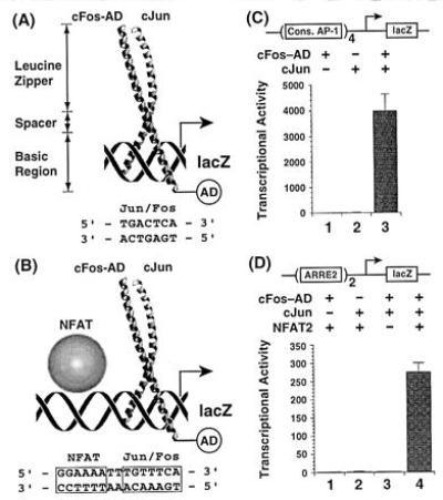
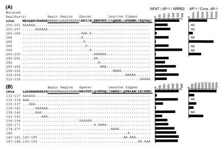
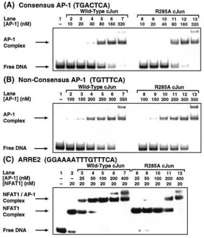
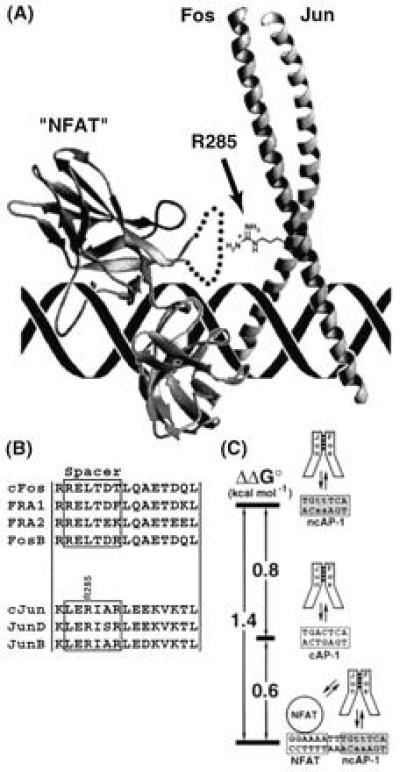
The heterologous transcription factors NFAT and AP-1 coordinately regulate cytokine gene expression through cooperative binding to precisely juxtaposed DNA recognition elements. The molecular origins of cooperativity in the binding of NFAT and AP-1 to DNA are poorly understood. Herein we have used yeast one-hybrid screening and alanine-scanning mutagenesis to identify residues in AP-1 that affect cooperative interactions with NFAT on DNA. Mutation of a single conserved Arg residue to Ala in the cJun spacer region (R285A) led to a virtually complete abolition of cooperative interactions with NFAT. The DNA-binding activity of AP-1 alone was unaffected by the cJun R285A mutation, thus indicating that this residue influences cooperative binding only. Ala-scanning mutations elsewhere in AP-1, including the cFos subunit, revealed no other strongly interacting single positions. We thus conclude that NFAT contacts AP-1 in the spacer region of the cJun subunit, making an especially important contact to R285, and that these interactions drive formation of the cooperative NFAT/AP-1/DNA complex. These results provide a general strategy for selectively ablating cooperativity between transcription factors without affecting their ability to act alone and yield insights into the structural basis for coordinate regulation of gene expression.


Figures

Figure 1
Figure 1
Activation of lacZ reporter gene transcription by AP-1 acting alone and in cooperation with NFAT. (A) Structure of the AP-1 bZIP domain (22), with functional domains denoted, bound to a consensus AP-1 site. (B) NFAT, bound to a polypurine tract in ARRE2, cooperatively recruits AP-1 to the adjacent nonconsensus AP-1 site, thereby activating reporter gene expression. AD denotes the B42 activation domain, which is fused to the basic region of Fos. (C) AP-1-driven reporter assay detects the binding of AP-1 alone to a consensus site in yeast. (D) ARRE2-linked reporter assay detects the reconstitution of the cooperative AP-1/NFAT/ARRE2 complex in yeast. Transcriptional activity refers to units of β-galactosidase activity.
Figure 2
Figure 2
Effects of alanine-scanning mutations on AP-1 activity in vivo. Sequence of the bZIP region of the AP-1 subunits cJun (A) and cFos (B), with positions mutated to alanine denoted below. To the right of the sequences are shown the transcriptional activity of wild-type and mutant AP-1 proteins in the ARRE2 reporter assay, and (for selected examples) in the AP-1 reporter assay. ND, not determined. The standard errors in the measurements of transcriptional activity assays are estimated at ±25%.
Figure 3
Figure 3
EMSA analysis of DNA-binding activity in vitro. (A and B) Wild-type AP-1 and R285A cJun mutant AP-1 bind with essentially identical affinities to a consensus AP-1 site (Kd = ≈80 nM) (A) and to the nonconsensus site in ARRE2 (Kd = ≈300 nM) (B). (C) The R285A cJun mutant AP-1 shows a substantially diminished ability to cooperate with NFAT1. From the data in C, the Kd of wild-type AP-1 for the NFAT1/ARRE2 complex is estimated at ≈30 nM.
Figure 4
Figure 4
Cooperative contacts between NFAT and AP-1. (A) Notional model of the NFAT/AP-1/DNA complex, constructed by docking the x-ray structure of AP-1 (22) in the correct orientation (21) with that of an NF-κB p50 RHR monomer (“NFAT”) (39). Because the insert region of p50 is not homologous to the corresponding sequence in NFAT, it was replaced in the model with a schematic loop (dotted line), part of which is believed to contact the DNA minor groove (L. Chen and G.L.V., unpublished results). The side chain of R285 in cJun, shown explicitly, projects toward NFAT. (B) Sequence alignments of mammalian Jun and Fos family members in the spacer region and first heptad repeat, which in Jun contacts NFAT. (C) Free-energy diagram illustrating the influence of sequence composition and NFAT cooperativity on the energetics of AP-1/DNA interactions. Stability of the complexes increases from top to bottom.

References

    1. Crabtree G R, Clipstone N A. Annu Rev Biochem. 1994;63:1045–1083. - PubMed
    1. Durand D B, Bush M R, Morgan J G, Weiss A, Crabtree G R. J Exp Med. 1987;165:395–407. - PMC - PubMed
    1. Shaw J-P, Utz P J, Durand D B, Toole J J, Emmel E A, Crabtree G R. Science. 1988;241:202–205. - PubMed
    1. Durand D B, Shaw J-P, Bush M R, Replogle R E, Belagaje R, Crabtree G R. Mol Cell Biol. 1988;8:1715–1724. - PMC - PubMed
    1. Rao A. Immunol Today. 1994;15:274–281. - PubMed

Publication types

MeSH terms

LinkOut - more resources