. 1996 Nov 26;93(24):13726-30.
doi: 10.1073/pnas.93.24.13726.

Defective subunit assembly underlies a digenic form of retinitis pigmentosa linked to mutations in peripherin/rds and rom-1

Affiliations

Defective subunit assembly underlies a digenic form of retinitis pigmentosa linked to mutations in peripherin/rds and rom-1

A F Goldberg et al. Proc Natl Acad Sci U S A. .

Abstract

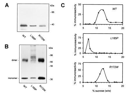
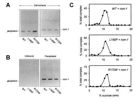
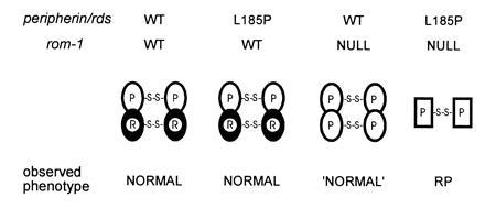
Retinitis pigmentosa (RP) is a group of progressive retinal dystrophies that include the most common hereditary degenerative disease affecting the retina. Although most disease phenotypes appear to result from defects at single genetic loci (monogenic), at least one instance of RP appears to require a coinheritance of defects in the unlinked peripherin/rds and rom-1 alleles (digenic), which encode the polypeptide subunits of an oligomeric transmembrane protein complex present at photoreceptor outer segment disc rims. Sedimentation velocity analysis was performed upon the affected gene products expressed heterologously in COS-1 cells to examine the assembly of the subunit polypeptides. The results indicate that the missense peripherin/rds mutant, L185P, which segregates with instance of digenically inherited RP, is conditionally defective with respect to its subunit assembly. Unlike wild-type peripherin/rds, the L185P mutant does not form native-like homotetramers on its own; however, the L185P mutant can assemble with wild-type rom-1 to form a structurally normal heterotetrameric complex. These findings provide a novel molecular-based rationale for the unusual digenic disease inheritance pattern and offer insight into regions of peripherin/rds and rom-1, which contribute to subunit-subunit interactions.


Figures

Figure 1
Figure 1
Expression and oligomerization of WT and mutant peripherin/rds. COS-1 cells transfected with the indicated expression plasmids (pcPER, pcR172W, or pcL185P) were detergent-solubilized 72 h posttransfection and probed by Western blot analysis with anti-peripherin/rds monoclonal antibody per2B6. Approximately 1.5 μg of total protein per lane was subjected to SDS/PAGE in the presence (A) or absence (B) of reducing agent (2% 2-mercaptoethanol). (C) Sedimentation velocity analysis of WT and mutant peripherin/rds complexes. Immunoaffinity-purified peripherin/rds variants from transfected COS-1 cells were sedimented through 5–20% sucrose gradients and sedimentation profiles were determined by chemiluminescent Western blotting (with anti-peripherin/rds per2B6) and laser densitometry as described. In the absence of rom-1, WT peripherin/rds is assembled as a homotetramer in COS-1 cells (32, 33).
Figure 2
Figure 2
Assembly of WT and mutant peripherin/rds–rom-1 oligomeric complexes. (A) Detergent extracts from COS-1 cells cotransfected with plasmids encoding rom-1 (6 μg, pcROM) and the indicated peripherin/rds variant (6 μg) were assayed for the expression of each protein by Western blot analysis with either anti-peripherin/rds monoclonal antibody per2B6 (Left) or anti-rom-1 polyclonal antibody romC2 (Right). (B) A sequential immunoprecipitation (anti-peripherin)/Western blot (anti-rom-1) analysis was performed to assay assembly of rom-1 with each of the peripherin/rds variants. Peripherin/rds was quantitatively immunoprecipitated; in each instance, no reactivity is seen in the unbound fractions (Left). Rom-1 is observed to coprecipitate in each instance indicating heteromeric assembly with each of the mutants (Right). (C) Sedimentation velocity analysis of WT and mutant peripherin/rds–rom-1 complexes. Peripherin/rds-containing complexes were immunoaffinity-purified from transfected COS-1 cells and sedimented through 5–20% sucrose gradients. Sedimentation profiles of assembled heteromeric complexes were determined by chemiluminescent Western blotting with an anti-rom-1 antibody and laser densitometry as described (33).
Figure 3
Figure 3
A molecular rationale for digenic RP involving the L185P peripherin/rds mutation and a rom-1 null mutation. This model stresses the functional importance of peripherin/rds-containing tetramers; instabilities (leading to RP disease phenotypes) are predicted in photoreceptors lacking sufficient levels of these particular species. Unaffected (WT) individuals and heterozygotes for a L185P mutation are expected to have normal levels of peripherin/rds–rom-1 heterotetramers, resulting in stable outer segments and an essentially normal phenotype. Heterozygotes for a null rom-1 mutation are expected to be borderline normal (“normal”) since peripherin/rds homotetramers can partially compensate for reduced levels of peripherin/rds–rom-1 heterotetramers. Double heterozygotes for the L185P peripherin/rds mutation and a null rom-1 mutation exhibit the RP phenotype. Reduced levels of the peripherin/rds–rom-1 heterotetramer coupled with the inability of L185P to form homotetramers will likely lead to disorganized and unstable outer segments.

References

    1. Heckenlively J R. Retinitis Pigmentosa. Philadelphia: Lippincott; 1988.
    1. Berson E L. Invest Ophthalmol Vis Sci. 1993;34:1659–1676. - PubMed
    1. Shastry B S. Am J Med Genet. 1994;52:467–474. - PubMed
    1. Daiger S P, Sullivan L S, Rodriguez J A. Behav Br Sci. 1995;18:452–467.
    1. Dryja T P, Li T. Hum Mol Genet. 1995;4:1739–1743. - PubMed

Publication types

MeSH terms

LinkOut - more resources