. 1996 Nov 26;93(24):13748-53.
doi: 10.1073/pnas.93.24.13748.

Molecular characterization of the human peroxisomal branched-chain acyl-CoA oxidase: cDNA cloning, chromosomal assignment, tissue distribution, and evidence for the absence of the protein in Zellweger syndrome

Affiliations

Molecular characterization of the human peroxisomal branched-chain acyl-CoA oxidase: cDNA cloning, chromosomal assignment, tissue distribution, and evidence for the absence of the protein in Zellweger syndrome

E Baumgart et al. Proc Natl Acad Sci U S A. .

Abstract

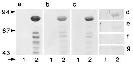
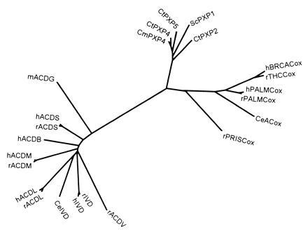
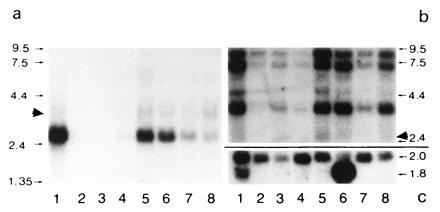
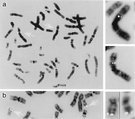
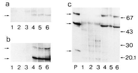
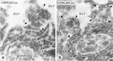
Peroxisomes in human liver contain two distinct acyl-CoA oxidases with different substrate specificities: (i) palmitoyl-CoA oxidase, oxidizing very long straight-chain fatty acids and eicosanoids, and (ii) a branched-chain acyl-CoA oxidase (hBRCACox), involved in the degradation of long branched fatty acids and bile acid intermediates. The accumulation of branched fatty acids and bile acid intermediates leads to severe mental retardation and death of the diseased children. In this study, we report the molecular characterization of the hBRCACox, a prerequisite for studying mutations in patients with a single enzyme deficiency. The composite cDNA sequence of hBRCACox, derived from overlapping clones isolated via immunoscreening and hybridization of human liver cDNA expression libraries, consisted of 2225 bases and contained an open reading frame of 2046 bases, encoding a protein of 681 amino acids with a calculated molecular mass of 76,739 Da. The C-terminal tripeptide of the protein is SKL, a known peroxisome targeting signal. Sequence comparison with the other acyl-CoA oxidases and evolutionary analysis revealed that, despite its broader substrate specificity, the hBRCACox is the human homolog of rat trihydroxycoprostanoyl-CoA oxidase (rTHCCox) and that separate gene duplication events led to the occurrence in mammals of acyl-CoA oxidases with different substrate specificities. Northern blot analysis demonstrated that--in contrast to the rTHCCox gene--the hBRCACox gene is transcribed also in extrahepatic tissues such as heart, kidney, skeletal muscle, and pancreas. The highest levels of the 2.6-kb mRNA were found in heart, followed by liver. The enzyme is encoded by a single-copy gene, which was assigned to chromosome 3p14.3 by fluorescent in situ hybridization. It was absent from livers of Zellweger patients as shown by immunoblot analysis and immunocytochemistry.


Figures

Figure 1
Figure 1
Nucleotide and deduced amino acid sequence of hBRCACox. The nucleotide sequence shown is derived from overlapping clones of two different libraries and numbered in 5′ → 3′ direction. Numbers on the left of the sequences indicate the appropriate nucleotide or amino acid position. Nucleotides in the 5′ leader sequence are indicated by negative numbers. The position of the stop codon is indicated by ∗∗∗. The amino acid sequences of tryptic peptides are underlined. Their experimentally determined masses agreed with their calculated masses. In addition, some other peptides could be positioned according to their mass [measured and calculated mass (M+H)+ given within parentheses]: amino acids 102–108 (871.2; 871.9), 109–120 (1265.2; 1266.4), 395–409 (1697.9; 1696), and 544–552 (1111.2; 1112.3).
Figure 2
Figure 2
Relationship between the different acyl-CoA oxidases visualized in Western blots. Aliquots of bacterial lysate (20 μg of protein), containing thioredoxin–hBRCACox fusion protein (theoretical molecular mass, 91 kDa), were analyzed for cross-reactivity with different antisera. (a) Anti-thioredoxin. (b) Anti-hBRCACox. (c) Anti-rTHCCox. (d) Anti-SKL. (e) Anti-rPALMCox (B subunit). (f) Anti-rPALMCox (C subunit). (g) Anti-rPRISCox. Lanes 1 and 2 represent a noninduced culture and an induced culture, respectively. Migration of molecular weight markers (expressed in kDa) is indicated at the left.
Figure 3
Figure 3
Evolutionary analysis of acyl-CoA oxidases and acyl-CoA dehydrogenases. Multiple sequence alignment of the protein sequences was performed using the pileup program (34) from the GCG package as well as with clustal w (35). The phylogenetic tree was constructed according to the neighbor-joining method (36), as implemented in the neighbor program while the distance matrices needed were calculated with protdist, both from the phylip package (37). The reliability of the tree was assessed by bootstrap analysis. Bootstrap values were in general higher than 80%. Whereas all known acyl-CoA oxidases were aligned, a selection of acyl-CoA dehydrogenases, possessing distinct substrate specificities, was made (data bank accession numbers are also given) as follows: hBRCACox/rTHCCox, X95189X95189; h/rPALMCox I, X71440X71440 and P07872P07872; rPRISCox, X95188X95188; CeACox (Caenorhabditis elegans acyl-CoA oxidase), P34355P34355; ScPXP1 (S. cerevisiae POX1 protein), P13711P13711; CtPXP2/4/5 (Candida tropicalis POX2/4/5 proteins), P06598P06598, P11356P11356, and P08790P08790; CmPXP4 (Candida maltosa POX4 protein), P05335P05335; mACDG (mouse glutaryl-CoA dehydrogenase), U18992U18992; h/rACDS (human and rat short-chain acyl-CoA dehydrogenases), P16219P16219 and P15651P15651; hACDB (human short branched-chain acyl-CoA dehydrogenase), P45954P45954; h/rACDM (human and rat medium-chain acyl-CoA dehydrogenases), P11310P11310 and P08503P08503; h/rACDL (human and rat long-chain acyl-CoA dehydrogenases), P28330P28330 and P15650P15650; rACDV (rat very-long-chain acyl-CoA dehydrogenase), P45953P45953; Ce/h/rIVD (C. elegans, human, and rat isovaleryl-CoA dehydrogenases), P34275P34275, P26440P26440, and P12007P12007. The h/rPALMCoxs II are not shown since the exon duplication, giving rise to the type I and II mRNAs, is a recent event and the branches are hardly visible. Similar topologies were seen in trees obtained by other programs, the tree-construction option in clustal w (35) or the quartet puzzling method (38) (data not shown).
Figure 4
Figure 4
Northern blot analysis of BRCACox in human tissues. A human multiple tissue blot (CLONTECH) containing ≈1 μg of purified mRNA of different tissues was analyzed for the presence of hBRCACox mRNA (a). Lanes: 1, heart; 2, brain; 3, placenta; 4, lung; 5, liver; 6, skeletal muscle; 7, kidney; 8, pancreas. After longer exposures, signals corresponding to BRCACox mRNA became also visible in lanes 2–4 (data not shown). For comparison, in b, the signals obtained for hPALMCox mRNA are shown, while the amount of β-actin mRNA is shown in c. Despite the stringent washing conditions, a very slight cross-reactivity between the mRNAs for hBRCACox and hPALMCox was noticed (marked by solid arrowheads). This is probably due to a stretch of bases with high homology in the middle portion of the mRNA sequences. The migration of standards (expressed in kb) are indicated on both sides (a and b). The values at the right of c correspond to the length (in kb) of the mRNAs for the distinct β-actin isoforms.
Figure 5
Figure 5
Chromosomal assignment of the hBRCACox gene. The chromosomal mapping of the hBRCACox gene was investigated by means of fluorescent in situ hybridization of metaphase spreads of white blood cells with biotinylated probes. Labeling was confined to chromosome 3p14.3 (a) and no cross-reactivity of the cDNA probe with the hPALMCox gene was found. In agreement with earlier reports (18), the latter gene was assigned to chromosome 17q25.1 (b). The signals, associated with the alleles of the oxidase genes on chromosomes 3 and 17 of the metaphase spreads are indicated by open arrows. The inserts at the right side show several examples of chromosomes 3 (a) and 17 (b) at a higher magnification, demonstrating specific hybridization of the probes with both alleles.
Figure 6
Figure 6
Western blot analysis of hBRCACox in liver of control subjects and Zellweger patients. Aliquots of liver homogenates (25 μg of protein) from different Zellweger patients (lanes 1–3) and healthy individuals (lanes 4–6) were subjected to SDS/PAGE followed by blotting (in triplicate). The blots were incubated with anti-hBRCACox (a), anti-rPALMCox (B subunit) (b), or anti-rPALMCox (A subunit) (c). Lane P in c represents 200 ng of purified rPALMCox. No specific signals for either hBRCACox or PALMCox protein were detectable in Zellweger patients. The small arrows at the left of a refer to the position of the 70-kDa hBRCACox subunit band, of b to the positions of the 70- and 51-kDa subunits of hPALMCox, and of c to the position of the 70-, 51-, and 23-kDa subunits of hPALMCox. Migration of the molecular mass standards (expressed in kDa) is indicated only at the right of c.
Figure 7
Figure 7
Immunocytochemical localization of BRCACox in human liver peroxisomes. Pieces of human liver were processed for immunoelectron microscopy and stained for hBRCACox (a) and hPALMCox (b). Peroxisomes (marked with arrowheads) are specifically labeled with gold particles depicting the different acyl-CoA oxidase antigens. Other cell organelles such as mitochondria (MITO), endoplasmic reticulum (ER), lysosomes, or nuclei were not labeled. Peroxisomes in this sample are often associated with glycogen deposits (GLY). The sample was derived from a ruptured and, therefore, nontransplanted liver lobe of a liver prepared for transplantation. Due to delay between excision of the liver and fixation, the sections show signs of autolysis. In contrast to the positive results (despite the autolysis) obtained with the liver of the healthy individual, no specific labeling was obtained in sections of a well-preserved liver biopsy of a Zellweger patient, either with antibodies against hBRCACox or with the different antibodies against rPALMCox (data not shown).

References

    1. Mannaerts, G. P. & Van Veldhoven, P. P. (1995) Ann. N.Y. Acad. Sci. 804, in press. - PubMed
    1. van den Bosch H, Schutgens R B H, Wanders R J A, Tager J. Annu Rev Biochem. 1992;61:157–197. - PubMed
    1. Reddy J K, Mannaerts G P. Annu Rev Nutr. 1994;14:343–370. - PubMed
    1. Casteels M, Schepers L, Van Veldhoven P P, Eyssen H J, Mannaerts G P. J Lipid Res. 1990;31:1865–1872. - PubMed
    1. Vanhove G F, Van Veldhoven P P, Fransen M, Denis S, Eyssen H J, Wanders R J A, Mannaerts G P. J Biol Chem. 1993;268:10335–10344. - PubMed

Publication types

MeSH terms