. 1996 Nov 26;93(24):13754-9.
doi: 10.1073/pnas.93.24.13754.

Metal binding properties and secondary structure of the zinc-binding domain of Nup475

Affiliations

Metal binding properties and secondary structure of the zinc-binding domain of Nup475

M T Worthington et al. Proc Natl Acad Sci U S A. .

Abstract

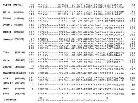
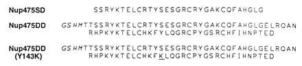
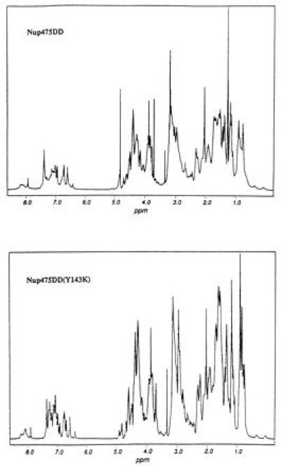
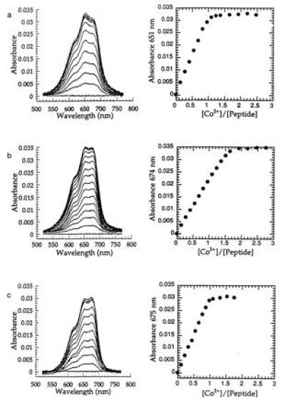
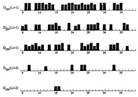
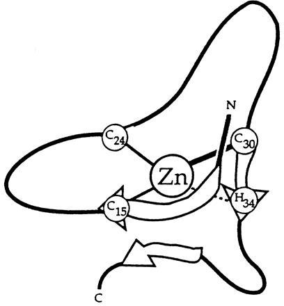
Nup475 is a nuclear zinc-binding protein of unknown function that is induced in mammalian cells by growth factor mitogens. Nup475 contains two tandemly repeated sequences YKTELCX8CX5CX3H (Cys3His repeats) that are thought to be zinc-bindin domains. Similar sequences have been found in a number of proteins from various species of eukaryotes. To determine the metal binding properties and secondary structure of the putative zinc-binding domains of Nup475, we have used synthetic or recombinant peptides that contain one or two domain sequences. The peptide with a single domain bound 1.0 +/- 0.1 equivalents of Co2+, and the peptide with two domains bound 1.7 +/- 0.4 equivalents of Co2+. Both peptides bound Co2+ and Zn2+ with affinities similar to those of classical zinc finger peptides. In each case, the Co2+ complex exhibited strong d-d transitions characteristic of tetrahedral coordination. For structural studies by nuclear magnetic resonance spectroscopy, we used a more soluble two-domain peptide that had a single amino acid substitution in a nonconserved amino acid residue in the second Cys3His repeat. The mutant peptide unexpectedly showed loss of one of its metal binding sites and displayed ordered structure for only the first Cys3His sequence. On the basis of the nuclear magnetic resonance data, we propose a structure for the Nup475 metal-binding domain in which the zinc ion is coordinated by the conserved cysteines and histidine, and the conserved YKTEL motif forms a parallel sheet-like structure with the C terminus of this domain. This structure is unlike that of any previously described class of metal binding domain.


Figures

Figure 1
Figure 1
Alignment of Nup475 Cys3His domain homologs. Nup475-related Cys3His domains were identified through a Genquest Q search of the Swiss Prot data base using the Cys3His protein sequences of Nup475 and those of known family members to probe the database. Note the conservation of cysteines, histidines, glycines, and aromatic residues. The first seven proteins are previously described members of this family, the last seven are the result of our database search. A striking feature of the latter group is the number of proteins known to be involved in some form of posttranslational RNA metabolism or as part of RNA viruses, although in no case has this motif been shown to directly bind RNA. Not included are the highly conserved human, rat, and bovine homology of Nup475/TTP/TIS11, TIS11b, and TIS11d; only the murine proteins are shown. (GenBank accession nos. for the proteins shown: Nup475, M58691M58691; TIS11b, M58566M58566; TIS11d, M58564M58564; YTIS11p, S76619S76619; DTIS11, U13397U13397; Unkempt, Z11527Z11527; zfs1+, D49913D49913; TIScc1, X81194X81194; human U2AF35, M96982M96982; human U2AFBP-RS, S69507S69507; Drosophila melanogaster Suppressor of sable, M57889M57889; human respiratory synticial virus A2, M11486M11486; turkey rhinotracheitis virus, X63408X63408; and bovine respiratory synticial virus, M82816M82816).
Figure 2
Figure 2
Sequences of the synthetic peptide Nup475SD and the recombinant, thrombin-cleaved wild-type Nup475DD and Nup475DD(Y143K) mutant two-domain peptides. Additional residues conferred by the pET28b vector in the two-domain proteins are italicized; the residue mutated from tyrosine-to-lysine in Nup475DD(Y143K) is underlined.
Figure 3
Figure 3
One-dimensional proton NMR in D2O of wild-type two-domain protein Nup475DD and the Nup475DD(Y143K) mutant. Line-broadening in case of the wild-type sample is consistent with aggregation. The comparable overall shape of the two spectra suggests that the protons are in similar chemical environment and are therefore in the same overall protein fold.
Figure 4
Figure 4
Absorption spectra of Co2+ complexes of proteins and peak absorbance vs. equivalents of metal bound of Nup475SD (a); Nup475DD (b); and Nup475DD(Y143K) (c). The Nup475SD spectra shown are at 43.9 μmol peptide, the Nup475DD spectra are at 26.2 μmol protein, and the Nup475(Y143K) spectra are at 30.1 μmol protein. The overall shapes of the d-d absorbances are similar to previously reported TFIIIA-type zinc fingers mutated to have Cys3His tetrahedral coordination (17). One of the effects of the Tyr to Lys mutation in Nup475(Y143K) (c) is that the mutant binds only one equivalent of metal, unlike the wild-type protein from which it was derived (b). Zinc back-titrations were performed in the same system using a 300-fold excess of cobalt.
Figure 5
Figure 5
Sequential NOEs for amino acids 9–41 of the Nup475(Y143K) peptide. Numbering refers to the cleaved recombinant peptide, with the first amino acid after the cleavage site numbered 1. The height of the column represents the relative intensity of the cross peak. Not shown are HNCACB connectivities that run the span of the entire segment shown.
Figure 6
Figure 6
Schematic of the proposed secondary structure of the first Nup475 domain. The central zinc atom is coordinated by the three spatially conserved cysteines and histidine. Note the two turns and the suggestion of a parallel sheet formed by the conserved Y10KTEL14 motif and the C30QFAH34 residues surrounding the coordinating histidine.

References

    1. Lai W S, Stumpo D J, Blackshear P J. J Biol Chem. 1990;265:16556–16563. - PubMed
    1. Varnum B C, Ma Q F, Chi T H, Fletcher B, Herschman H R. Mol Cell Biol. 1991;11:1754–1758. - PMC - PubMed
    1. Dubois R N, McLane M W, Ryder K, Lau L F, Nathans D. J Biol Chem. 1990;265:19185–19191. - PubMed
    1. Varnum B C, Lim R W, Kujubu D A, Luner S J, Kaufman S E, Greenberger J S, Gasson J C, Herschman H R. Mol Cell Biol. 1989;9:3580–3583. - PMC - PubMed
    1. Dubois R N, Bishop P R, Graves-Deal R, Coffey R J. Cell Growth Differ. 1995;6:523–529. - PubMed

Publication types

MeSH terms

LinkOut - more resources