. 1996 Nov 26;93(24):13760-5.
doi: 10.1073/pnas.93.24.13760.

The Saccharomyces CDC13 protein is a single-strand TG1-3 telomeric DNA-binding protein in vitro that affects telomere behavior in vivo

Affiliations

The Saccharomyces CDC13 protein is a single-strand TG1-3 telomeric DNA-binding protein in vitro that affects telomere behavior in vivo

J J Lin et al. Proc Natl Acad Sci U S A. .

Abstract

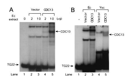
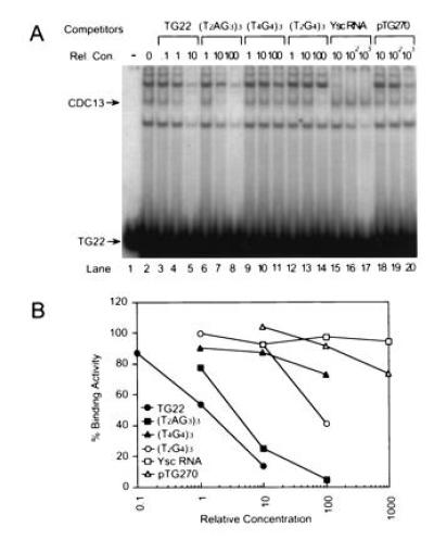
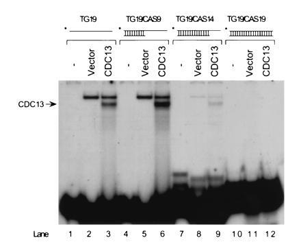
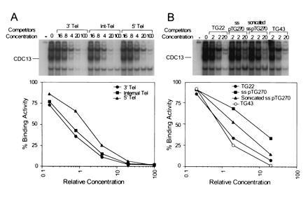
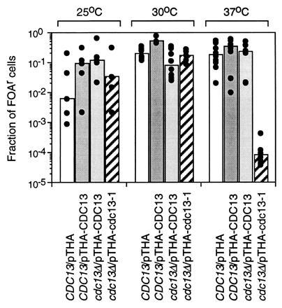
Saccharomyces telomeres consist of approximately 300 bp of C1-3A/TG1-3 DNA. Cells lacking the activity of the essential gene CDC13 display a cell cycle arrest mediated by the DNA damage sensing, RAD9 cell cycle checkpoint, presumably because they exhibit strand-specific loss of telomeric and telomere-adjacent DNA [Garvik, B., Carson, M. & Hartwell, L. (1995) Mol. Celi. Biol. 15,6128-6138]. Cdc13p expressed in Escherichia coli or overexpressed in yeast bound specifically to single-strand TG1-3 DNA. The specificity of binding displayed by Cdc13p in vitro indicates that in vivo it could bind to both the short, constitutive single-strand TG1-3 tails thought to be present at telomeres at most times in the cell cycle as well as to the long single-strand TG1-3 tails that are intermediates in telomere replication. Genes located near yeast telomeres are transcriptionally repressed, a phenomenon known as telomere position effect. Cells overexpressing a mutant form of Cdc13p had reduced telomere position effect at high temperatures. These data suggest that Cdc13p functions by binding directly to telomeric DNA, thereby limiting its accessibility to degradation and transcription as well as masking it from factors that detect damaged DNA.


Figures

Figure 1
Figure 1
Cdc13p is a ss TG1–3 binding protein. 32P-labeled TG22, a 22-base oligonucleotide of ss TG1–3 DNA (Table 1), was used in gel mobility-shift assays using total extracts from either bacteria (Ec) or yeast (Ysc). All gel mobility-shift assays reported here were done in the presence of ss poly(dI·dC). E. coli extracts were prepared from cells expressing Cdc13p or carrying the vector alone. Yeast extracts were prepared from a CDC13 strain carrying vector alone or pTHA-CDC13 for overexpression of Cdc13p. The arrows point to a TG22-protein complex that was seen only in E. coli cells expressing Cdc13p or yeast cells overexpressing Cdc13p. For both panels, the first lane had no extract. (A) The amount of extract used is indicated. (B) Two micrograms of extract was used.
Figure 2
Figure 2
Cdc13p binding is specific. (A) Gel mobility-shift assays were carried out using 1 ng of 32P-labeled TG22 and 10 μg of E. coli extract from cells expressing Cdc13p. Prior to loading the gel, the TG22 was mixed with unlabeled ss oligonucleotides of the sequence and at the relative concentration (Rel. Con.) indicated above each lane or with total yeast RNA or pTG270, a 6300-bp plasmid containing 270 bp of C1–3A/TG1–3 DNA. The Cdc13p–TG22 complex is indicated by the arrow. Lane 1 had no extract. (B) The amount of gel shift activity in the Cdc13p-TG22 complex was quantified for each oligonucleotide at each concentration. Relative concentration of competitor nucleic acids in all gels was expressed as the ratios of the amount of ss TG1–3 DNA in the competitor to the amount of ss TG1–3 DNA in TG22 (see Materials and Methods); for competitors that did not have ss TG1–3 DNA (e.g., Ysc RNA), it was expressed as the ratio of nanograms of competitor to nanograms of TG22. In this and subsequent gels, the amount of gel shifted complex in TG22 with no competitor was defined as 100%. The values presented are the average of two independent experiments; for each point, the two values were within 14% of each other.
Figure 3
Figure 3
Cdc13p binds tailed duplex molecules. Gel mobility-shift assays were carried out using 10 μg of extract from E. coli cells expressing Cdc13p or carrying vector alone. 32P-labeled TG19 was annealed to various oligonucleotides to generate substrates with 9 bp of duplex DNA and a 10-base ss TG1–3 tail (TG19/CAS9), with 14 bp of duplex DNA and a 5-base ss TG1–3 tail (TG19/CAS14), or duplex TG19 (TG19/CAS19). The arrow indicates the position of the Cdc13p-DNA complex. The complex migrating more slowly than the Cdc13p-DNA complex in the vector-alone lanes was present in reduced amounts in lane 8, compared with lanes 2 and 5, and absent in lane 11 because it was due to an E. coli-encoded ss TG1–3 DNA binding activity, and there was less (lane 8) or no (lane 11) ss TG1–3 DNA in the DNA substrates used in those lanes.
Figure 4
Figure 4
Cdc13p binding does not require an end. Gel mobility-shift assays were carried out using 1 ng of 32P-labeled TG22 and 10 μg of extract from E. coli expressing Cdc13p. TG22 was mixed with unlabeled competitor DNA, extract was added, and the samples were examined by electrophoresis. The % binding activity was determined as described in Fig. 2. In the interest of space, only the top portion of each gel is shown. (A) The competitor DNAs were unlabeled 42-base ss oligonucleotides having TG22 at its 3′ end (3′ Tel), in the middle of the oligonucleotide (Internal Tel), or at its 5′ end (5′ Tel). (B) The competitor DNA was unlabeled TG22, ss pTG270, a circular 6300-base ss phagemid containing 276 bases of ss TG1–3 DNA, ss pTG270 sheared to an average size of 1000 bases, or TG43, an oligonucleotide having 43 bases of ss TG1–3 DNA.
Figure 5
Figure 5
TPE is reduced at high temperature in cells overexpressing Cdc13-1p. CDC13 or cdc13Δ cells with URA3 next to the telomere of chromosome VII-L and carrying the plasmids indicated below the bars were spread on plates containing or lacking FOA and incubated at the indicated temperatures. The top of the bar is the median value for the fraction of FOAR cells, with dots indicating values for independent colonies. Independent colonies with identical values are represented by a single dot.

References

    1. Zakian V A. Science. 1995;270:1601–1607. - PubMed
    1. Shampay J, Szostak J W, Blackburn E H. Nature (London) 1984;310:154–157. - PubMed
    1. Oka Y, Shiota S, Nakai S, Nishida Y, Okubo S. Gene. 1980;10:301–306. - PubMed
    1. Klobutcher L A, Swanton M T, Donini P, Prescott D M. Proc Natl Acad Sci USA. 1981;78:3015–3019. - PMC - PubMed
    1. Henderson E R, Blackburn E H. Mol Cell Biol. 1989;9:345–348. - PMC - PubMed

Publication types

MeSH terms