. 1996 Nov 26;93(24):13786-90.
doi: 10.1073/pnas.93.24.13786.

Inhibition of interleukin 1 beta-converting enzyme-mediated apoptosis of mammalian cells by baculovirus IAP

Affiliations

Inhibition of interleukin 1 beta-converting enzyme-mediated apoptosis of mammalian cells by baculovirus IAP

C J Hawkins et al. Proc Natl Acad Sci U S A. .

Abstract

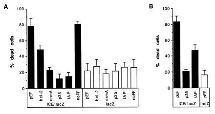
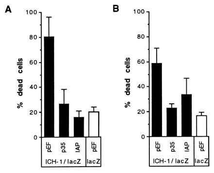
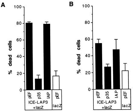
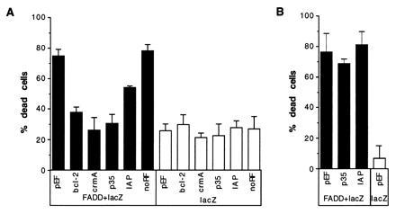
Apoptosis can be a potent weapon against viral infection and consequently has selected for viruses carrying antiapoptosis genes. Two baculovirus proteins, IAP and p35, can prevent insect cells from dying in response to infection. p35, which interferes with members of the Ced-3 family of cysteine proteases, can also function in mammalian cells. We investigated the ability of IAP from Orgyia pseudotsugata nuclear polyhedrosis virus to prevent death of mammalian cells. IAP was transiently expressed in mammalian cells and its ability to block cell death caused by expression of interleukin-1 beta converting enzyme (ICE), FADD, or the ICE homologues ICH-1 and ICE-Lap3, was investigated. IAP strongly inhibited ICE- and ICH-1-induced cell death but protected only partially against death by overexpression of FADD and not at all against death due to enforced ICE-Lap3 expression. These results demonstrate that a baculoviral IAP protein can functionally interact with conserved components of the apoptosis machinery in mammalian cells.


Figures

Figure 1
Figure 1
ICE-mediated apoptosis of HeLa cells. X-Gal-stained HeLa cells transiently transfected with the following plasmids: lacZ (a), ICE/lacZ (b and c), and ICE/lacZ (d and e) with pEF-OpIAP. Cells were scored as dead or alive based upon their appearance by phase contrast (Left) or conventional (Right) microscopy, as described by Miura et al. (5). Live cells (arrows without tails) were large and flat with an irregular shape and smooth edges. Dead cells (arrows with tails) were small, refractile, and round, with irregular “blebbed” membranes. Some had detached from the culture dish.
Figure 2
Figure 2
Inhibition of ICE-mediated cell death by IAP expression. (A) HeLa cells were transiently cotransfected with a p32ICE-lacZ fusion plasmid and with expression plasmids carrying either no insert, bcl-2, crmA, p35, OpIAP, or truncated IAP lacking the sequences encoding the RING finger (designated noRF). Parallel wells of HeLa cells were cotransfected with the same test plasmids and a lacZ plasmid. At least five cultures were transfected and counted blind for each test plasmid; as for all figures, values indicate the percentage of X-Gal-stained cells that were dead (mean ± 2 SEM) for each set of replicates. (B) CHO cells transfected with lacZ plasmid and empty pEF vector (lane 1) and cotransfections of ICE/lacZ and either pEF, p35, or OpIAP expression plasmids. Three transfections of CHO cells were performed for each test plasmid.
Figure 3
Figure 3
Protection from ICH-1-induced death by OpIAP. HeLa (A) or CHO (B) cells were cotransfected with the ICH-1/lacZ plasmid and empty vector, p35, or OpIAP expression constructs. A cotransfection in which the lacZ plasmid and pEF vector were introduced into cells reflects the background death due to transfection (bar on the left panel).
Figure 4
Figure 4
Inability of IAP to prevent death due to expression of ICE-Lap3. Cells, either 293T (A) or CHO (B) were cotransfected with ICE-Lap3 p28 plasmid, lacZ, and either empty vector or a plasmid bearing p35 or IAP. Two days after the addition of serum after transfection, the cells were stained and the blue (transfected) cells were scored for viability. At least three wells for each plasmid combination were transfected and scored blind.
Figure 5
Figure 5
Partial suppression of death due to FADD expression by OpIAP. Plasmids expressing Bcl-2, CrmA, p35, IAP, only the amino-terminal half of OpIAP, or the empty vector were cotransfected into HeLa cells with a lacZ plasmid and a FADD expression plasmid (A). Parallel control wells were also transfected with the test plasmids and lacZ plasmid. At least five wells for each test plasmid were transfected and scored blind. The empty vector, p35, and OpIAP plasmids were also cotransfected with the lacZ and FADD plasmids into CHO cells in the same manner as the HeLa transfection (B).

References

    1. Vaux D L, Häcker G, Strasser A. Cell. 1994;76:777–779. - PubMed
    1. Steller H. Science. 1995;267:1445–1462. - PubMed
    1. Kumar S. Trends Biochem Sci. 1995;20:198–202. - PubMed
    1. Yuan J, Shaham S, Ledoux S, Ellis H M, Horvitz H R. Cell. 1993;75:641–652. - PubMed
    1. Miura M, Zhu H, Rotello R, Hartweig E A, Yuan J. Cell. 1993;75:653–660. - PubMed

Publication types

MeSH terms