. 1996 Nov 26;93(24):13902-7.
doi: 10.1073/pnas.93.24.13902.

Cell cycle-regulated generation of single-stranded G-rich DNA in the absence of telomerase

Affiliations

Cell cycle-regulated generation of single-stranded G-rich DNA in the absence of telomerase

I Dionne et al. Proc Natl Acad Sci U S A. .

Abstract

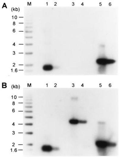
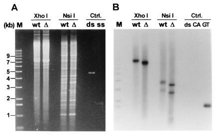
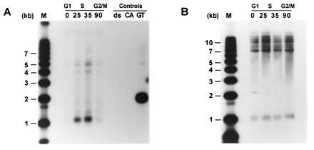
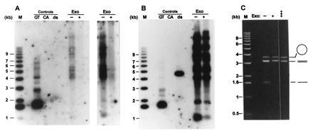
Current models of telomere replication predict that due to the properties of the polymerases implicated in semiconservative replication of linear DNA, the two daughter molecules have one end that is blunt and one end with a short 3' overhang. Telomerase is thought to extend the short 3' overhang to produce long single-stranded overhangs. Recently, such overhangs, or TG1-3 tails, were shown to occur on both telomeres of replicated linear plasmids in yeast. Moreover, indirect evidence suggested that the TG1-3 tails also occurred in a yeast strain lacking telomerase. We report herein a novel in-gel hybridization technique to probe telomeres for single-stranded DNA. Using this method, it is shown directly that in yeast strains lacking the TLC1 gene encoding the yeast telomerase RNA, TG1-3 single-stranded DNA was generated on chromosomal and plasmid telomeres. The single-stranded DNA only appeared in S phase and was sensitive to digestion with a single-strand-specific exonuclease. These data demonstrate that during replication of telomeres, TG1-3 tails can be generated in a way that is independent of telomerase-mediated strand elongation. In wild-type strains, these TG1-3 tails could subsequently serve as substrates for telomerase and telomere binding proteins on all telomeres.


Figures

Figure 1
Figure 1
Nondenaturing in-gel hybridization technique to detect single-stranded TG1–3 DNA. In both gels, lanes 1 and 2 contained, respectively, 4 ng and 0.4 ng of single-stranded phagemid DNA derived from pGT75; lanes 3 and 4 contained, respectively, 30 ng and 3 ng of linearized double-stranded pMW75 DNA; and lanes 5 and 6 contained, respectively, 30 ng and 3 ng of linearized pMW75 DNA that was heat-denatured for 10 min at 95°C prior to loading. M denotes end-labeled ladder DNA. The gels were treated for in-gel hybridization. The gel in A was treated as a nondenaturing gel, whereas the gel in B was denatured in the gel prior to hybridization to the end-labeled CA-oligo probe.
Figure 2
Figure 2
Analysis of the telomeres of the ≈7.5-kb linear plasmid YLpFAT10. Low molecular weight DNA was extracted via a Hirt procedure from TLC1+ (lanes marked wt) and tlc1Δ (lanes marked Δ) cells. The DNA was digested with the indicated enzymes and analyzed on a 0.75% agarose gel. There are no recognition sites for XhoI on YLpFAT10, leaving the plasmid intact in those lanes. There are two recognition sites for NsiI, yielding three fragments for YLpFAT10: the two terminal fragments are ≈2.5 kb and ≈3.5 kb and the internal fragment is ≈1.5 kb (22). (A) The gel stained with ethidium bromide. (B) An autoradiograph of the same gel after hybridization to the end-labeled CA-oligo. Lane M contains end-labeled molecular weight marker DNA. The DNAs loaded under the heading Ctrl (Controls) were as follows: ds, linearized double-stranded pMW75 DNA; CA and ss, single-stranded phagemid DNA derived from pCA75; GT, single-stranded phagemid DNA derived from pGT75.
Figure 3
Figure 3
Single-stranded TG1–3 DNA appears in a cell cycle-controlled manner on chromosomal telomeres of tlc1Δ cells. Total DNA was isolated from yeast strain RWY14 (tlc1::LEU2) at the following time points: 0, cells at 37°C were arrested at START due to the temperature-sensitive cdc7 gene; 25, 35, and 90, cells were released into a synchronous S phase for 25, 35, or 90 min, respectively. The control DNA loadings (Controls) were as in Fig. 2B. M, molecular weight standards. Above the actual time points, the corresponding approximate cell cycle phases are indicated. The DNAs were digested with the restriction enzyme XhoI and separated on a 0.7% agarose gel. The gel in A was treated as a nondenaturing gel and hybridized to the end-labeled CA-oligo. The gel in B was identical to the gel in A except that this gel was then treated as a regular Southern blot (i.e., the DNA was denatured in the gel) using a Y′-specific probe.
Figure 4
Figure 4
The single-stranded TG1–3 DNAs detected on telomeres of tlc1Δ cells are TG1–3 tails. Total yeast DNA isolated from RWY14 cells was incubated either without (−) or with (+) E. coli exonuclease I for 10 min at 37°C. The DNAs were then precipitated and digested with the restriction endonuclease XhoI. The DNA loadings under Controls are as in Fig. 2. M, molecular weight standards. The gel in A was treated as a nondenaturing gel and probed with the CA-oligo. Two exposures of the lanes containing the genomic DNAs are shown to emphasize the absence of detectable bands in the lane with the exonuclease-treated DNA. After appropriate exposures of the gel in A were obtained, the DNA in the gel was denatured in 0.5 M NaOH and rehybridized to the CA-oligo. (B) An autoradiogram of the rehybridized gel. Note that the double-stranded control DNA now is detected and that the pattern of TG1–3 repeat containing bands is the same in the exonuclease-treated and untreated DNA. (C) The exonuclease used in this experiment displays the expected specificity. Single-stranded circular DNA (M13mp18 phage DNA, open circle), linearized double-stranded pVZ1 DNA (3.2 kb, double lines), and linear single-stranded DNA (heat-denatured pVZ1 DNA, single line) were mixed and incubated in the absence of the exonuclease (−) or in the presence of exonuclease for 10 min (+) or 45 min (+++). The DNAs were then separated on a 0.6% agarose gel and stained with ethidium bromide. Note that the linear single-stranded DNA was completely degraded after 10 min of incubation, while the amount of circular single-stranded M13 DNA remained virtually unchanged even after 45 min of incubation.

References

    1. Blackburn E H. Nature (London) 1991;350:569–573. - PubMed
    1. Zakian V A. Science. 1995;270:1601–1607. - PubMed
    1. Shampay J, Szostak J W, Blackburn E H. Nature (London) 1984;310:154–157. - PubMed
    1. Klobutcher L A, Swanton M T, Donini P, Prescott D M. Proc Natl Acad Sci USA. 1981;78:3015–3019. - PMC - PubMed
    1. Pluta A F, Kaine B P, Spear B B. Nucleic Acids Res. 1982;10:8145–8154. - PMC - PubMed

Publication types

LinkOut - more resources