

Costimulatory function and expression of CD40 ligand, CD80, and CD86 in vascularized murine cardiac allograft rejection
- PMID: 8943044
- PMCID: PMC19478
- DOI: 10.1073/pnas.93.24.13967
Costimulatory function and expression of CD40 ligand, CD80, and CD86 in vascularized murine cardiac allograft rejection
Abstract
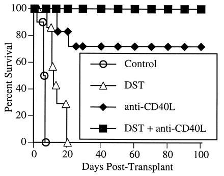
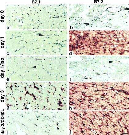
Recent data implicates a role for the CD40-CD40 ligand (CD40L) pathway in graft rejection. One potential mechanism is direct costimulation of T cells through CD40L. Alternatively, the ability of CD40 stimulation to induce CD80 (B7-1) and CD86 (B7-2) expression on antigen-presenting cells (APCs) has led to the hypothesis that the role of CD40-CD40L interactions in transplant rejection might be indirect, i.e., to promote the costimulatory capacity of APCs. Here, we have used a murine vascularized cardiac allograft model to test this hypothesis. Treatment of the recipients with donor splenocytes and a single dose of anti-CD40L mAb induces long-term graft survival (> 100 days) in all animals. This is associated with marked inhibition of intragraft Th1 cytokine [interferon gamma and interleukin (IL) 2] and IL-12 expression with reciprocal up-regulation of Th2 cytokines (IL-4 and IL-10). In untreated allograft recipients, CD86 is strongly expressed on endothelial cells and infiltrating mononuclear cells of the graft within 24 hr. In contrast, CD80 expression is not seen until 72 hr after engraftment. Anti-CD40L mAb has no detectable effect on CD86 up-regulation, but almost completely abolishes induction of CD80. However, animals treated with anti-CD80 mAb or with a mutated form of CTLA4Ig (which does not bind to CD86) rejected their cardiac allografts, indicating that blockade of CD80 alone does not mediate the graft-prolonging effects of anti-CD40L mAb. These data support the notion that the role of CD40-CD40L in transplant rejection is not solely to promote CD80 or CD86 expression, but rather that this pathway can directly and independently costimulate T cells. These data also suggest that long-term graft survival can be achieved without blockade of either T cell receptor-mediated signals or CD28-CD86 engagement.
Figures

References
-
- Janeway C H, Bottomly K. Cell. 1994;76:275–285. - PubMed
-
- Schwartz R H. Cell. 1989;57:1073–1081. - PubMed
-
- Guinan E C, Gribben J G, Boussiotis V A, Freeman G J, Nadler L M. Blood. 1994;84:3261–3282. - PubMed
-
- June C H, Bluestone J A, Nadler L M, Thompson C B. Immunol Today. 1994;15:321–331. - PubMed
-
- Sayegh M H, Turka L A. J Am Soc Nephrol. 1995;6:1143–1150. - PubMed
Publication types
MeSH terms
Substances
Grants and funding
LinkOut - more resources
Full Text Sources
Other Literature Sources
Medical
Molecular Biology Databases
Research Materials
Miscellaneous
