. 1996 Nov 26;93(24):13973-8.
doi: 10.1073/pnas.93.24.13973.

The tumor necrosis factor receptor 2 signal transducers TRAF2 and c-IAP1 are components of the tumor necrosis factor receptor 1 signaling complex

Affiliations

The tumor necrosis factor receptor 2 signal transducers TRAF2 and c-IAP1 are components of the tumor necrosis factor receptor 1 signaling complex

H B Shu et al. Proc Natl Acad Sci U S A. .

Abstract

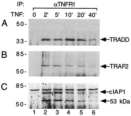
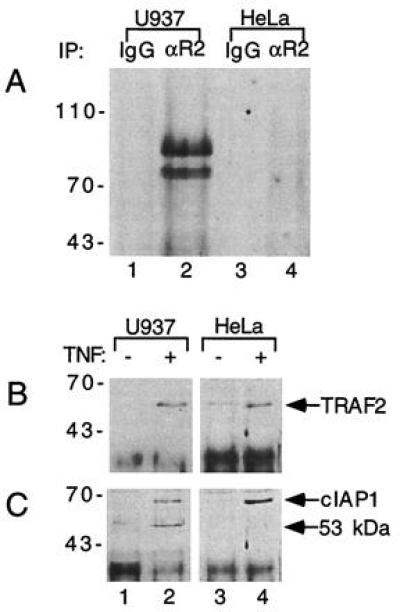
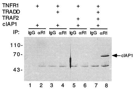
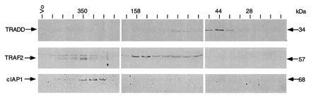
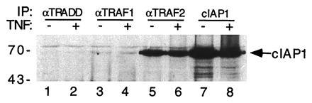
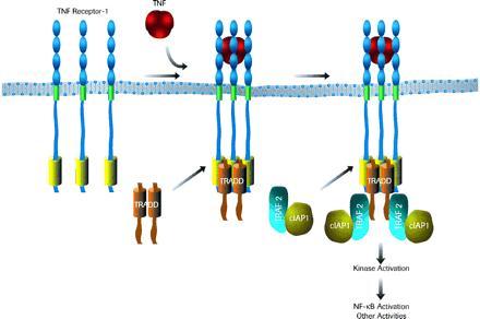
The two cell surface receptors for tumor necrosis factor (TNF) interact with a number of intracellular signal transducing proteins. The association of TRADD, a 34-kDa cytoplasmic protein containing a C-terminal death domain, with aggregated TNF receptor 1 (TNF-R1) through their respective death domains leads to NF-kappa B activation and programmed cell death. In contrast, TNF receptor 2 (TNF-R2) interacts with the TNF receptor associated factors 2/1 (TRAF2/TRAF1) heterocomplex, which mediates the recruitment of two cellular inhibitor of apoptosis proteins (c-IAP1 and c-IAP2) to TNF-R2. Here we show that the TNF-R2 signal transducers TRAF2 and c-IAP1 are a part of the TNF-R1 signaling complex. The recruitment of TRAF2 and c-IAP1 to TNF-R1 is TNF-dependent, is mediated by TRADD, and is independent of TNF-R2. These data establish the physiological involvement of TRAF2 and c-IAP1 in TNF-R1 signaling and help provide a molecular explanation for both the overlapping and distinct signals generated by the two TNF receptors.


Figures

Figure 1
Figure 1
TNF-dependent recruitment of TRADD, TRAF2, and c-IAP1 to TNF-R1 in U937 cells. TNF (100 ng/ml) was used to treat U937 cells (≈2 × 10⁸) for the indicated times (lanes 2–6) or left the U937 cells were left untreated (lane 1). Immunoprecipitations were carried out with the 985 mAb to TNF-R1. Immunoprecipitates were analyzed by immunoblotting with rabbit polyclonal antibodies against TRADD (A), TRAF2 (B), or c-IAP1 (C). Positions of molecular mass standards (in kDa) are shown on the left.
Figure 2
Figure 2
TNF-dependent recruitment of TRAF2 and c-IAP1 to TNF-R1 is TNF-R2-independent. (A) Comparison of TNF-R2 expression in U937 and HeLa cells. Lysates of U937 cells (≈1 × 10⁸) or HeLa cells (≈1 × 10⁸) were immunoprecipitated with the 1040 mAb to TNF-R2 (lanes 2 and 4) or mouse IgG control (lanes 1 and 3). The immunoprecipitates were subsequently analyzed by immunoblotting with a polyclonal TNF-R2 antibody. Positions of molecular mass standards (in kDa) are shown on the left. (B and C) Recruitment of TRAF2 and c-IAP1 to the TNF-R1 complex in U937 and HeLa cells. TNF (100 ng/ml) was used to treat 2 × 10⁸ U937 cells (lanes 1 and 2) or HeLa cells (lanes 3 and 4) for 5 min (lanes 2 and 4). Lanes 1 and 3 were left untreated. Cell lysates were immunoprecipitated with the 985 mAb to TNF-R1 and immunoprecipitates were analyzed by immunoblotting with polyclonal antibodies against TRAF2 (B) or c-IAP1 (C). Positions of molecular mass standards (in kDa) are shown on the left.
Figure 3
Figure 3
TRADD and TRAF2 mediate recruitment of c-IAP1 to TNF-R1. Combinations of expression vectors for TNF-R1, myc tagged c-IAP1, TRADD, and TRAF2 (as indicated) were used to transfect 293 cells (≈2 × 10⁶). Transfected cell lysates were immunoprecipitated with either mouse IgG control (lanes 1, 3, 5, and 7) or a goat anti human TNF-R1 antibody (lanes 2, 4, 6, and 8). Immunoprecipitates were analyzed for c-IAP1 by immunoblotting with a myc epitope antibody. Positions of molecular mass standards (in kDa) are shown on the left.
Figure 4
Figure 4
Biochemical properties of TRADD, TRAF2, and c-IAP1 in HeLa cells. HeLa cell lysates (1 × 10⁸ cells) were separated by size exclusion chromatography on Superdex 200 column. Individual fractions were analyzed by immunoblotting with polyclonal antibodies against TRADD, TRAF2, or c-IAP1 as indicated. Positions of molecular mass standards (in kDa) are shown on the left.
Figure 5
Figure 5
Association of TRAF2 and c-IAP1 in HeLa cells. HeLa cells (≈2 × 10⁸) were treated with TNF (100 ng/ml) for 5 min (lanes 2, 4, 6, and 8) or left untreated (lanes 1, 3, 5, and 7). Cell lysates were immunoprecipitated with rabbit polyclonal antisera against TRADD (lanes 1 and 2), TRAF1 (lanes 3 and 4), TRAF2 (lanes 5 and 6), or c-IAP1 (lanes 7 and 8). Immunoprecipitates were analyzed by immunoblotting with a polyclonal antibody against c-IAP1. Positions of molecular mass standards (in kDa) are shown on the left.
Figure 6
Figure 6
A model for the assembly of TNF-R1 signaling complex. See text for details.

References

    1. Goeddel D V, Aggarwal B B, Gray P W, Leung D W, Nedwin G E, Palladino M A, Patton J S, Pennica D, Shepard H M, Sugarman B J, Wong G H W. Cold Spring Harbor Symp Quant Biol. 1986;51:597–609. - PubMed
    1. Beutler B, Cerami A. Annu Rev Biochem. 1988;57:505–518. - PubMed
    1. Fiers W. FEBS Lett. 1991;285:199–212. - PubMed
    1. Loetscher H, Pan Y-C E, Lahm H W, Gentz R, Brockhaus M, Tabuch H, Lesslauer W. Cell. 1990;61:351–359. - PubMed
    1. Schall T J, Lewis M, Koller K J, Lee A, Rice G C, Wong G H W, Gatanaga T, Granger G A, Lentz R, Raab H, Kohr W J, Goeddel D V. Cell. 1990;61:361–370. - PubMed

MeSH terms

LinkOut - more resources