. 1996 Nov 26;93(24):14014-9.
doi: 10.1073/pnas.93.24.14014.

A novel mechanism of action of tetracyclines: effects on nitric oxide synthases

Affiliations

A novel mechanism of action of tetracyclines: effects on nitric oxide synthases

A R Amin et al. Proc Natl Acad Sci U S A. .

Abstract

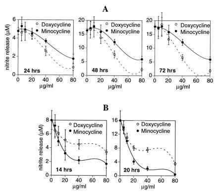
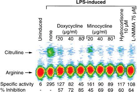
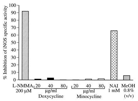
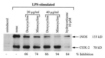
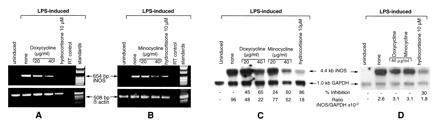
Tetracyclines have recently been shown to have "chondroprotective" effects in inflammatory arthritides in animal models. Since nitric oxide (NO) is spontaneously released from human cartilage affected by osteoarthritis (OA) or rheumatoid arthritis in quantities sufficient to cause cartilage damage, we evaluated the effect of tetracyclines on the expression and function of human OA-affected nitric oxide synthase (OA-NOS) and rodent inducible NOS (iNOS). Among the tetracycline group of compounds, doxycycline > minocycline blocked and reversed both spontaneous and interleukin 1 beta-induced OA-NOS activity in ex vivo conditions. Similarly, minocycline > or = doxycycline inhibited both lipopolysaccharide- and interferon-gamma-stimulated iNOS in RAW 264.7 cells in vitro, as assessed by nitrite accumulation. Although both these enzyme isoforms could be inhibited by doxycycline and minocycline, their susceptibility to each of these drugs was distinct. Unlike acetylating agents or competitive inhibitors of L-arginine that directly inhibit the specific activity of NOS, doxycycline or minocycline has no significant effect on the specific activity of iNOS in cell-free extracts. The mechanism of action of these drugs on murine iNOS expression was found to be, at least in part, at the level of RNA expression and translation of the enzyme, which would account for the decreased iNOS protein and activity of the enzyme. Tetracyclines had no significant effect on the levels of mRNA for beta-actin and glyceraldehyde-3-phosphate dehydrogenase nor on levels of protein of beta-actin and cyclooxygenase 2 expression. These studies indicate that a novel mechanism of action of tetracyclines is to inhibit the expression of NOS. Since the overproduction of NO has been implicated in the pathogenesis of arthritis, as well as other inflammatory diseases, these observations suggest that tetracyclines should be evaluated as potential therapeutic modulators of NO for various pathological conditions.


Figures

Figure 1
Figure 1
(A) Effect of doxycycline and minocycline on OA-NOS expression in human OA-affected cartilage. OA-affected knee articular cartilage from one OA-affected patient was placed in organ culture in 2 ml of medium in the presence or absence (control) of doxycycline and minocycline at 5–80 μg/ml. The spontaneous release of nitrite was monitored at different time intervals. Data are expressed as micromolar nitrite released (mean ± SD, n = 3–4). Zero time indicates spontaneous release of NO at 24, 48, and 72 h, which was 4.8 ± 0.38, 16.4 ± 0.7, and 17.8 ± 0.9, respectively. Statistics were derived using unpaired Student’s t test. Data represent one of three identical experiments with samples from different patients. (B) Effect of doxycycline and minocycline on nitrite accumulation in RAW 264.7 cells stimulated with LPS. Murine macrophage cells (RAW 264.7) were incubated with doxycycline or minocycline (5–80 μg/ml) for 1–2 h followed by addition of LPS at 100 ng/ml. After 14–20 h of incubation, the medium was used to estimate nitrite accumulation by the modified Griess reaction (23). Data are expressed as micromolar of nitrite accumulated at a given time interval (n = 3). Statistics were derived using unpaired Student’s t test. Data represent one of three similar experiments. Graphs were plotted using the delta graph curve-fitting program: polynomial of degree 3.
Figure 2
Figure 2
Effect of doxycycline and minocycline on iNOS enzyme activity in RAW 264.7 cells stimulated with LPS. Murine macrophage cells (RAW 264.7) were incubated with doxycycline or minocycline (20–80 μg/ml), hydrocortisone (10 μM), or l-NMMA (75 μM) for 1 h, followed by addition of LPS at 100 ng/ml for 16–18 h. The cell-free extracts were prepared and the l-arginine → l-citrulline conversion assay was carried out in cell-free extracts. The data have been represented as specific activity of the enzyme, which was defined as pmol of citrulline per min per mg of protein. Percent inhibition represents comparison with the LPS-stimulated cells in the absence of any modulator. Data represent one of three similar experiments.
Figure 3
Figure 3
Effect of doxycycline and minocycline on the specific activity of iNOS in vitro. RAW 264.7 cells were stimulated with LPS for 16 h and cell-free extracts were prepared as a source of iNOS. Various modulators including methanol (MeOH), the carrier for N-acetylimidazole (NAI), were added 15 min prior to the addition of the cofactors to initiate the iNOS reaction. The specific activity was calculated and percent inhibition compared with the original LPS-stimulated extract; 100% specific activity was 200 pmol of citrulline per min per mg of protein. Data represent one of two similar experiments.
Figure 4
Figure 4
Western blot analysis of iNOS in RAW 264.7 cells exposed to doxycycline or minocycline in the presence of LPS. RAW 264.7 cells activated with LPS (100 ng/ml) for 16–18 h, with and without doxycycline or minocycline (20–40 μg/ml) and hydrocortisone (10 μM) were analyzed for total iNOS and COX-2 protein. Briefly, 30 μg of cell extract was loaded onto SDS/PAGE gels and the filter probed simultaneously with anti-iNOS and anti-COX-2 mouse mAb followed with an anti-mouse serum conjugated to horseradish peroxidase on the same filters. Bands were quantitated on a densitometer and the percent inhibition was compared with the LPS-stimulated cells. Data represent one of four similar experiments.
Figure 5
Figure 5
Analysis of iNOS mRNA expression by RT–PCR and Northern blot in the presence and absence of tetracyclines. (A and B) RT–PCR analysis of iNOS and β-actin mRNA expression in RAW 264.7 cells was carried out after stimulation with LPS ± doxycycline, minocycline, or hydrocortisone. Equal amounts of RNA were analyzed for iNOS and β-actin expression. “RT control” designates preparation of RT–PCRs in the absence of reverse transcriptase using the LPS-stimulated RNA as the template from RAW 264.7 cells. Data represent one of two similar and separate experiments. (C and D) Northern blot analysis of iNOS and GAPDH mRNA expression in RAW 264.7 cells was carried out after stimulation with LPS ± doxycycline, minocycline, or hydrocortisone at 16 h (C) or 4 h (D) after stimulation. The iNOS/GAPDH signal was determined and quantitated using both a PhosphoImager and a densitometer. The percent inhibition of iNOS expression was normalized with the GAPDH signal and compared with the values of the LPS-stimulated cells. Data represent one of three similar experiments.

References

    1. Vane J R, Mitchell J A, Appleton I, Tomlinson A, Bishop-Bailey D, Croxtall J, Willoughby D A. Proc Natl Acad Sci USA. 1994;91:2046–2050. - PMC - PubMed
    1. Schmidt H H H W, Walter U. Cell. 1994;78:919–925. - PubMed
    1. Nathan C, Xie Q. Cell. 1994;78:915–918. - PubMed
    1. Marletta M A. Cell. 1994;78:927–930. - PubMed
    1. McCartney-Francis N, Allen J B, Mizel D E, Albina J E, Xie Q W, Nathan C F, Wahl S M. J Exp Med. 1993;178:749–754. - PMC - PubMed