Comparative Study
. 1996 Nov 26;93(24):14053-8.
doi: 10.1073/pnas.93.24.14053.

Tumor necrosis factor receptor-associated factor (TRAF)-1, TRAF-2, and TRAF-3 interact in vivo with the CD30 cytoplasmic domain; TRAF-2 mediates CD30-induced nuclear factor kappa B activation

Affiliations
Comparative Study

Tumor necrosis factor receptor-associated factor (TRAF)-1, TRAF-2, and TRAF-3 interact in vivo with the CD30 cytoplasmic domain; TRAF-2 mediates CD30-induced nuclear factor kappa B activation

S Ansieau et al. Proc Natl Acad Sci U S A. .

Retraction in

Abstract

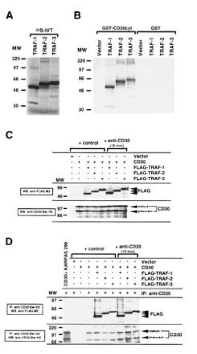
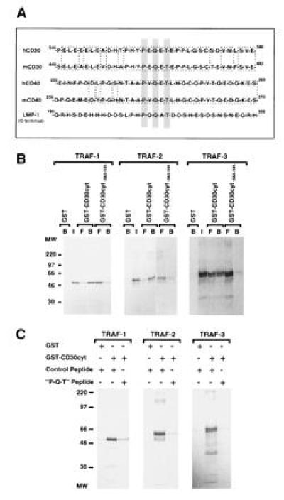
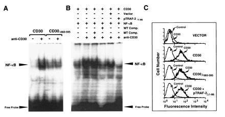
CD30 is a member of the tumor necrosis factor receptor superfamily, which can transduce signals for proliferation, death, or nuclear factor kappa B (NF-kappa B) activation. Investigation of CD30 signaling pathways using a yeast two-hybrid interaction system trapped a cDNA encoding the tumor necrosis factor receptor-associated factor (TRAF)-2 TRAF homology domain. TRAF-1 and TRAF-3 also interacted with CD30, and > 90% of in vitro-translated TRAF-1 or -2, or 50% of TRAF-3, bound to the CD30 cytoplasmic domain. TRAF-1, -2, and -3 bound mostly, but not exclusively, to the carboxyl-terminal 36 residues of CD30. The binding was strongly inhibited by a CD30 oligopeptide centered around a PXQXT (where X is any amino acid) motif shared with CD40 and the Epstein-Barr virus transforming protein LMP1, indicating that this motif in CD30 is an important determinant of TRAF-1, -2 or -3 interaction. At least 15% of TRAF-1, -2, or -3 associated with CD30 when coexpressed in 293 cells. The association was not affected by CD30 cross-linking. However, cross-linking of CD30 activated NF-kappa B. NF-kappa B activation was dependent on the carboxyl-terminal 36 amino acids of CD30 that mediate TRAF association. TRAF-2 has been previously shown to have a unique role in TRAF-mediated NF-kappa B activation, and NF-kappa B activation following CD30 cross-linking was blocked by a dominant negative TRAF-2 mutant. These data indicate that CD30 cross-linking-induced NF-kappa B activation is predominantly TRAF-2-mediated.


Figures

Figure 1
Figure 1
Association of human TRAF-1, TRAF-2, and TRAF-3 with the cytoplasmic domain of CD30. (A) TRAF-1, -2, and -3 proteins were [35S]methionine-labeled by in vitro translation (IVT). Ten microliters (20%) of each in vitro translation reaction was loaded. (B) The cytoplasmic domain of CD30 was fused to GST (GST–CD30cyt) and bound to glutathione beads. GST or GST–CD30cyt were incubated with 25% of the in vitro-translated TRAF proteins. The fraction bound to glutathione beads was analyzed by SDS/PAGE. Comparable GST loading was confirmed by staining with amino black and Western blotting against GST (data not shown). More than 90% of TRAF-1 and TRAF-2 and 50–60% of TRAF-3 respectively bound to GST–CD30cyt. (C) Western blot analysis of CD30 and FLAG expression in transfected 293 cells. 293 cells were transiently cotransfected with the full-length CD30 expression vector (5 μg) and the indicated FLAG-tagged TRAF constructs (5 μg). After 48 hr, lysates were prepared from unstimulated and anti-CD30-stimulated (15 min) transfected 293 cells. Thirty microliters of lysates (1/30) were separated by SDS/PAGE, blotted, preblocked, and probed for CD30 and/or FLAG-tagged TRAF proteins using anti-CD30 Ber-H2 or anti-FLAG M2 mAb followed by horseradish peroxidase-conjugated goat anti-mouse IgG and detection using enhanced chemiluminescence (FLAG indicates the transfected FLAG-tagged TRAF protein). The molecular weight (MW) standard is shown (in thousands). (D) In vivo interaction of TRAF-1, -2, and -3 with the cytoplasmic domain of CD30. One hundred microliters of the cell lysates analyzed in C were subjected to immunoprecipitation with the anti-CD30 Ber-H2 mAb. This mAb shows no cross-reactivity with the transfected TRAF proteins (data not shown). Coprecipitating FLAG-tagged TRAF proteins were detected by immunoblotting using the anti-FLAG M2 mAb (indicated by FLAG). KARPAS 299 is a large cell anaplastic lymphoma line.
Figure 2
Figure 2
A critical CD30 receptor sequence motif involved in TRAF binding. (A) A common sequence motif is involved in TRAF binding and is shared by CD30, CD40, and LMP1. Sequence aligment of the cytoplasmic domains of CD30, CD40, and LMP1 show the TRAF binding motif centered around the consensus sequence PXQXT (gray box). The mouse and human CD30 and CD40 sequences are highly conserved around the putative TRAF binding motif (1, 42, 43, 44). (B) The distal region of the cytoplasmic domain of CD30 is involved in TRAF binding. GST or GST-fusion proteins containing the full-length cytoplasmic domain of CD30 (CD30cyt) or a carboxyl-terminal deletion (CD30 lacking the carboxyl-terminal 36 aa; CD30cytΔ560-595) bound to glutathione beads were incubated with in vitro-translated TRAF-1, -2, and -3 proteins (25%), respectively. Reaction mixtures were analyzed by SDS/PAGE and autoradiography. A proximal deletion mutant of the cytoplasmic domain of CD30 (CD30cytΔ408-430) showed similar interaction as the full-length GST–CD30cyt fusion protein with all three TRAF proteins (data not shown). Western blotting confirmed that equal amounts of GST were used (data not shown). I, input; B, bound; and F, free. (C) Interaction of TRAF proteins with the cytoplasmic domain of CD30 was blocked by a peptide overlapping the TRAF binding domain in CD30. [35S]Methionine-labeled TRAF-1, -2, and -3 proteins (25%) were preincubated with 50-fold excess of a random control peptide (PSTMVYDACRMIRERIPEA) or the “P-Q-T” peptide encompassing amino acids 556–570 of the cytoplasmic domain of CD30 (HTPHYPEQETEPPLG) for 2 hr at 4°C, followed by incubation with the full-length cytoplasmic domain of CD30 fused to GST or GST alone for 1 hr at 4°C and analyzed by SDS/PAGE. Equal GST protein loading was confirmed by Western blotting (data not shown).
Figure 3
Figure 3
NF-κB activation of CD30-stimulated cells is mediated through TRAF proteins. (A) 293 cells were transfected with either the full-length CD30 cDNA or a deletion mutant lacking the distal 35 aa (CD30Δ560-595). Forty-eight hours after transfection, 293 cells were cross-linked using the anti-CD30 mAb M67 in the presence of a rabbit anti-mouse IgG1-specific antibody for 2 hr. Nuclear proteins were prepared and gel mobility shift assays were performed using a double-stranded oligodeoxynucleotide containing a NF-κB binding site as a probe. (B) 293 cells were cotransfected with the full-length CD30 expression vector (CD30) and either a control plasmid (Vector) or the negative dominant TRAF-2 plasmid (pTRAF-2Δ1-86). Forty-eight hours after transfection, 293 cells were cross-linked using the anti-CD30 mAb M67 plus rabbit anti-mouse IgG1-specific antibody (anti-CD30) for 2 hr. Nuclear proteins were prepared and analyzed for NF-κB activation by electrophoretic mobility-shift assay. Specificity of the complex was confirmed by competition assays using 50-fold molar excess of unlabeled NF-κB binding oligodesoxynucleotide (WT Comp.) or a mutated NF-κB site (MT Comp.). (C) CD30 surface expression of transfected 293 cells was determined by flow cytometry using the fluorescein isothiocyanate (FITC)-conjugated anti-CD30 mAb Ber-H2.

References

    1. Dürkop H, Latza U, Hummel M, Eitelbach F, Seed B, Stein H. Cell. 1992;68:421–427. - PubMed
    1. Smith C A, Farrah T, Goodwin R G. Cell. 1994;75:959–962. - PubMed
    1. Falini B, Pileri S, Pizzolo G, Dürkop H, Flenghi L, Stirpe F, Martelli M F, Stein H. Blood. 1995;85:1–14. - PubMed
    1. Schwab U, Stein H, Gerdes J, Lemke H, Kirchner H, Schaadt M, Diehl V. Nature (London) 1982;299:65–67. - PubMed
    1. Ellis T M, Simms P E, Slivnick D J, Jäck H-M, Fisher R I. J Immunol. 1993;151:2380–2389. - PubMed

Publication types

MeSH terms