. 1996 Nov 26;93(24):14128-33.
doi: 10.1073/pnas.93.24.14128.

Potential brain neuronal targets for amphetamine-, methylphenidate-, and modafinil-induced wakefulness, evidenced by c-fos immunocytochemistry in the cat

Affiliations

Potential brain neuronal targets for amphetamine-, methylphenidate-, and modafinil-induced wakefulness, evidenced by c-fos immunocytochemistry in the cat

J S Lin et al. Proc Natl Acad Sci U S A. .

Abstract

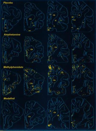
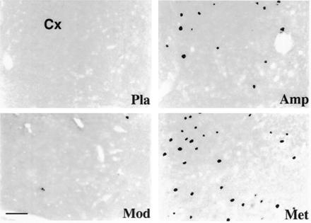
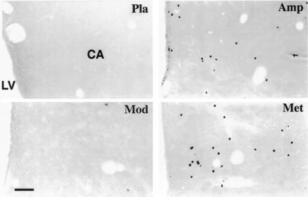
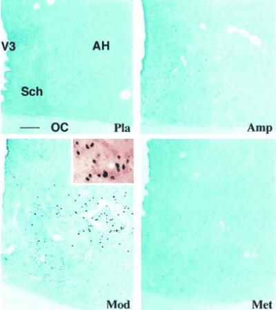
Much experimental and clinical data suggest that the pharmacological profile of modafinil, a newly discovered waking substance, differs from those of amphetamine and methylphenidate, two classical psychostimulants. The brain targets on which modafinil acts to induce wakefulness, however, remain unknown. A double-blind study using the protooncogene c-fos as experimental marker in the cat was, therefore, carried out to identify the potential target neurons of modafinil and compare them with those for amphetamine and methylphenidate. Cats were sacrificed after a single oral administration of amphetamine, methylphenidate, or modafinil at equivalent doses for wake induction (1, 2.5, or 5 mg/kg, respectively) and brain sections examined for Fos by immunocytochemistry. Administration of either amphetamine or methylphenidate evoked Fos-like immunoreactivity in a large number of neurons in the striatum and whole cortex, especially in the caudate nucleus and mediofrontal cortex, which are known to be dopaminergic targets. In contrast, administration of modafinil resulted in the labeling of few cells in these structures, but did induce marked Fos labeling in neurons of the anterior hypothalamic nucleus and adjacent areas. These results provide evidence for the potential brain targets of modafinil, which differ from those of amphetamine or methylphenidate, and suggest that modafinil induces wakefulness by mechanisms distinct from those of the two stimulants.


Figures

Figure 1
Figure 1
Distribution of Fos-like immunoreactivity in the rostral brain of the cat. Camera lucida drawing of frontal sections showing Fos-ir neurons following oral administration of the indicated substances Note that (i) the placebo induces little labeling, (ii) amphetamine or methylphenidate treatment causes numerous Fos-positive neurons in the cerebral cortex and striatum, and (iii) modafinil treatment induces little labeling of the cortex and striatum, but a large number of aggregated positive neurons is seen in the anterior hypothalamic nucleus (AH). ACC, nucleus accumbens; CA, caudate nucleus; CL, claustrum; DBH, diagonal band of Broca; GP, globus pallidus; IC, internal capsule; LV, lateral ventricle; OC, optic chiasma; OLT, olfactory tubercle; OT, optic tract; Para, anterior paraventricular nucleus of the thalamus; PrF, prefrontal cortex; Pu, putamen; SI, substantia innominata; V3, third ventricle.
Figure 2
Figure 2
Photomicrographs of frontal sections through the mediofrontal cortex (Cx) of the cat, showing Fos-like immunoreactivity following different administrations. Note that (i) the large number of stained cells seen with methylphenidate (Met) treatment and, to a lesser degree, with amphetamine (Amp) and (ii) few or no labeled neurons seen with modafinil (Mod) or placebo (Pla). (Bar = 50 μm.)
Figure 3
Figure 3
Photomicrographs of frontal sections through the caudate nucleus (CA) of the cat, showing Fos-like immunoreactivity following different administrations. Note the large number of stained cells seen with amphetamine (Amp) or methylphenidate (Met) treatment and that few or no labeled neurons are seen using modafinil (Mod) or placebo (Pla). LV, lateral ventricle. (Bar = 80 μm.)
Figure 4
Figure 4
Photomicrographs (blue filter) of frontal sections through the mediocaudal part of the preoptic/anterior hypothalamus of the cat, showing Fos-like immunoreactivity following different administrations. Note that (i) no obvious labeling is seen with placebo (Pla), (ii) some weakly labeled cells are present in the periventricular zone of the third ventricle (V3) following amphetamine (Amp) or methylphenidate (Met) treatment, and (iii) modafinil (Mod) induces some weakly stained neurons in the suprachiasmatic nucleus (Sch) and many strongly labeled cells in the AH. A higher power magnification of the Inset shown in the AH appears in the upper right photomicrograph (without filter), illustrating the morphology of the Fos-ir labeling in the cell nuclei on counterstained background. OC, optic chiasma. (Bar = 50 μm.)

References

    1. Bastuji H, Jouvet M. Prog Neuropsychopharmacol Biol Psychiatry. 1988;12:695–700. - PubMed
    1. Duteil J, Rambert F A, Pessonnier J, Hermant J F, Gombert R, Assous E. Eur J Pharmacol. 1990;180:49–58. - PubMed
    1. Hermant J F, Rambert F A, Duteil J. Psychopharmacology. 1991;103:28–32. - PubMed
    1. Lin J S, Roussel B, Akaoka H, Fort P, Debilly G, Jouvet M. Brain Res. 1992;591:319–326. - PubMed
    1. Morgan J I, Curran T. Trends Neurosci. 1989;12:459–462. - PubMed

Publication types

MeSH terms

LinkOut - more resources