. 1996 Dec 10;93(25):14355-60.
doi: 10.1073/pnas.93.25.14355.

Three-dimensional structure of human electron transfer flavoprotein to 2.1-A resolution

Affiliations

Three-dimensional structure of human electron transfer flavoprotein to 2.1-A resolution

D L Roberts et al. Proc Natl Acad Sci U S A. .

Abstract

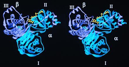
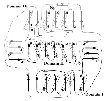
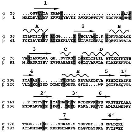
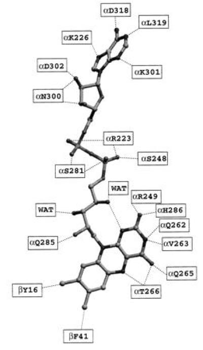
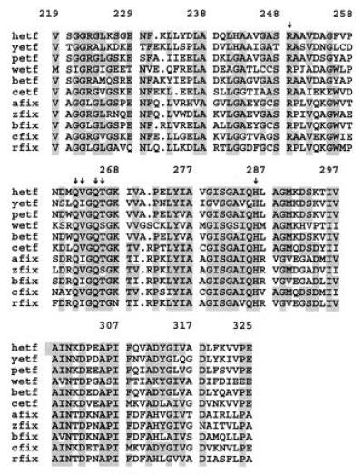
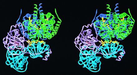
Mammalian electron transfer flavoproteins (ETF) are heterodimers containing a single equivalent of flavin adenine dinucleotide (FAD). They function as electron shuttles between primary flavoprotein dehydrogenases involved in mitochondrial fatty acid and amino acid catabolism and the membrane-bound electron transfer flavoprotein ubiquinone oxidoreductase. The structure of human ETF solved to 2.1-A resolution reveals that the ETF molecule is comprised of three distinct domains: two domains are contributed by the alpha subunit and the third domain is made up entirely by the beta subunit. The N-terminal portion of the alpha subunit and the majority of the beta subunit have identical polypeptide folds, in the absence of any sequence homology. FAD lies in a cleft between the two subunits, with most of the FAD molecule residing in the C-terminal portion of the alpha subunit. Alignment of all the known sequences for the ETF alpha subunits together with the putative FixB gene product shows that the residues directly involved in FAD binding are conserved. A hydrogen bond is formed between the N5 of the FAD isoalloxazine ring and the hydroxyl side chain of alpha T266, suggesting why the pathogenic mutation, alpha T266M, affects ETF activity in patients with glutaric acidemia type II. Hydrogen bonds between the 4'-hydroxyl of the ribityl chain of FAD and N1 of the isoalloxazine ring, and between alpha H286 and the C2-carbonyl oxygen of the isoalloxazine ring, may play a role in the stabilization of the anionic semiquinone. With the known structure of medium chain acyl-CoA dehydrogenase, we hypothesize a possible structure for docking the two proteins.


Figures

Figure 1
Figure 1
A stereo ribbon diagram of the human ETF structure. FAD (yellow) binds in a cleft formed by the α (blue) and β (lavender) subunits. The AMP (green) is located entirely within the β subunit. All ribbon models were made with the program ribbons (29).
Figure 2
Figure 2
Topology of the ETF protein, indicating a pseudo 2-fold between domains I and III. The α subunit (black), makes up domain I and domain IIα, and the β subunit (gray) consists of domains III and IIβ. α-helices (cylinders), β-strands (arrows), and random coils (lines) are indicated. Broken arrows indicate regions belonging to domain II, whereas solid arrows denote domains I and III.
Figure 3
Figure 3
Sequence alignment of domains I and III. The sequence alignment was made based on structural superposition of the two domains. Arrows above the sequence denote β-strands, whereas curves represent α-helices. The secondary structure elements are identified as denoted in Fig. 2. The top lines (α) represent domain I, whereas the bottom lines (β) are from domain III. Residues that are identical between the two domains are shaded. There is a 13.6% residue identity between the two domains, with an rms deviation of 2.1 Å between the Cα atoms of residues located either on helices or strands.
Figure 4
Figure 4
Residues involved in binding the FAD cofactor of ETF. Dotted lines indicate residues (or solvent) that are within hydrogen bonding distance (<3.3 Å) of one another. WAT denotes ordered water molecules. FAD is shown as it is modeled in ETF.
Figure 5
Figure 5
Sequence alignment of residues involved in binding FAD among members of the ETF α subunit family of proteins. Proteins used in the sequence alignment are: hetf, human ETF (32); petf, P. denitrificans ETF (33); yetf, Saccharomyces cerevisiae hypothetical ETF α subunit; wetf, Methylophilus methylotrophus W3A1 ETF (9); betf, Bradyrhizobium japonicum putative ETF (GenBank accession no. U32230U32230); cetf, Clostridium acetobutylicum putative ETF (34); afix, Azorhizobium caulinodans putative FixB (35); zfix, Azotobacter vinelandii putative FixB, (Protein Identification Resource accession no. S49188S49188); bfix, B. japonicum putative FixB (36); cfix, C. acetobutylicum putative FixB (GenBank accession no. M91817M91817); and rfix, Rhizobium meliloti putative FixB (37). Residues that are identical in at least six of the sequences are shaded. Arrows indicate residues involved in binding the isoalloxazine portion of the FAD, as shown in Fig. 4.
Figure 6
Figure 6
A stereo ribbon diagram depicting the hypothetical docking of ETF to an MCAD dimer. The refined structures of human ETF and porcine MCAD (43) were used. The MCAD dimer (green and dark blue, mainly the green monomer) fits into the groove formed by the α (cyan) and β (lavender) subunits of ETF, in a manner close to the FAD of MCAD. By this mechanism, the closest flavin–flavin distance is approximately 19.5 Å.

References

    1. Crane F L, Beinert H. J Biol Chem. 1956;218:717–731. - PubMed
    1. Beinert H. Enzymes. 1963;7:467–476.
    1. Ikeda Y, Tanaka K. J Biol Chem. 1983;258:1077–1085. - PubMed
    1. Leinich A C, Goodman S I. J Biol Chem. 1986;261:4090–4096. - PubMed
    1. Ikeda Y, Tanaka K. J Biol Chem. 1983;258:9477–9487. - PubMed

Publication types

Associated data