. 1996 Dec 10;93(25):14373-8.
doi: 10.1073/pnas.93.25.14373.

Crystal structure of the breakpoint cluster region-homology domain from phosphoinositide 3-kinase p85 alpha subunit

Affiliations

Crystal structure of the breakpoint cluster region-homology domain from phosphoinositide 3-kinase p85 alpha subunit

A Musacchio et al. Proc Natl Acad Sci U S A. .

Abstract

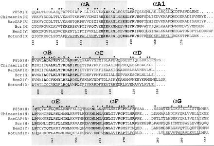
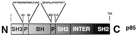
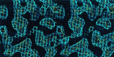
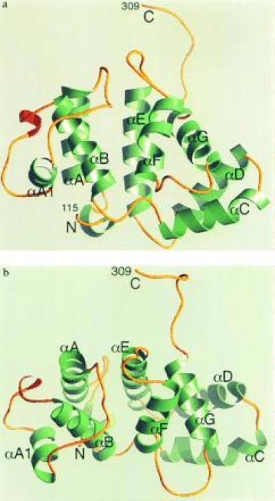
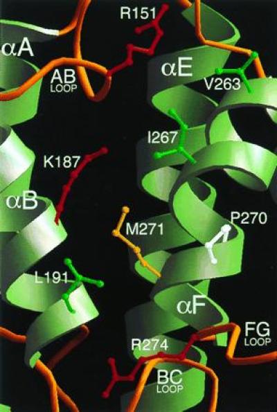
Proteins such as the product of the break-point cluster region, chimaerin, and the Src homology 3-binding protein 3BP1, are GTPase activating proteins (GAPs) for members of the Rho subfamily of small GTP-binding proteins (G proteins or GTPases). A 200-residue region, named the breakpoint cluster region-homology (BH) domain, is responsible for the GAP activity. We describe here the crystal structure of the BH domain from the p85 subunit of phosphatidylinositol 3-kinase at 2.0 A resolution. The domain is composed of seven helices, having a previously unobserved arrangement. A core of four helices contains most residues that are conserved in the BH family. Their packing suggests the location of a G-protein binding site. This structure of a GAP-like domain for small GTP-binding proteins provides a framework for analyzing the function of this class of molecules.


Figures

Figure 1
Figure 1
A sequence alignment of a subset of BRC-homology (BH) domains. A larger alignment, comprising 22 sequences, was used to evaluate the degree of sequence conservation at each position. The sequences used, with their database accession number for each sequence listed last in parentheses, are reported (B, bovine; CE, Caenorhabditis elegans; D, Drosophila melanogaster; H, human; M, mouse; R, rat; Y, Saccharomyces cerevisiae): (H) breakpoint cluster region (Bcr) (P11274); (H) ABR (U01147); (M) 3BP1 (X87671); (R) p122 (D31962); (R) p190 (M94721); (H) C1 (X78817); (Y) Bem2 (P39960); (Y) Bem3 (P32873); (R) β-chimaerin (Q03070); (H) IT5P (P32019); (Y) RGA1 (P39083); (D) Rotund (P40809); (M) p85α (P26450); (H) p85α ((P27986); (B) p85α (P23727); (B) p85β (P23726); (H) n-chimaerin (P15882); (R) n-chimaerin (P30337); (H) OCRL (Q01968); (Y) YB9G (P38339); (H) RhoGAP (Z23024); (CE) CeGAP (U02289). Residues that were conserved in at least 14 sequences out of 22 are shown in bold type. Asterisks above columns indicate residues whose side chains are part of the hydrophobic core of the molecule. Open circles identify residues that are part of the proposed ligand-binding site. Filled rectangles indicate residues lying at intron–exon boundaries in the mouse p85α gene (12). Each helical fragment boxed. The N- and C-terminal residues of each helix were defined as the first and last residues whose main chain carbonyl and amide groups, respectively, were involved in an intrahelical hydrogen bond. The gray boxes identify three blocks of conservation discussed in the text. Numbering corresponds to full-length human p85α. The alignment was generated with the pileup program (GCG package). The image was created using the programs Adobe illustrator and Adobe photoshop (Adobe Systems, Mountain View, CA).
Figure 2
Figure 2
Schematic view of the domain organization of p85. SH2 and SH3, Src-homology regions 2 and 3, respectively; P, proline-rich regions (see text); INTER, the inter-SH2 region that mediates dimerization with p110.
Figure 3
Figure 3
Stereo view of the averaged electron density map (contoured at 1 σ), shown against the final model. The map was used to build the model of the BH domain.
Figure 4
Figure 4
(a) Ribbon diagram of the PI3K BH domain. Molecule B is shown. α-Helices are in green, and 310 helices are in red. N and C indicate the N and C termini of the domain, respectively. The figure was created with the program ribbons (29). (b) A view of the domain from the top of the four-helix bundle, roughly corresponding to a 90° rotation about a horizontal axis parallel to the plane of the page in a.
Figure 5
Figure 5
The proposed G protein-binding site of the BH domain. The domain is viewed from the same orientation shown in Fig. 4A. Residues Arg-151, Lys-187, Pro-270, and Arg-274 are conserved in the BH family. Leu-191 and Val-263 are nonconserved residues occurring at sites that are otherwise strongly conserved (Fig. 1). The side chains of Ile-267 and Met-271 are included to show the relatively hydrophobic character of this patch.

References

    1. Bourne H R, Sanders D A, McCormick F. Nature (London) 1991;349:117–127. - PubMed
    1. Ridley A J. J Cell Sci Suppl. 1994;18:127–131. - PubMed
    1. Hall A. Cell. 1992;69:389–391. - PubMed
    1. Vojtek A B, Cooper J A. Cell. 1995;82:527–529. - PubMed
    1. Boguski M S, McCormick F. Nature (London) 1993;366:643–654. - PubMed

Publication types

MeSH terms

Substances