. 1996 Dec 10;93(25):14452-5.
doi: 10.1073/pnas.93.25.14452.

Dominant negative inhibition by fragments of a monomeric enzyme

Affiliations

Dominant negative inhibition by fragments of a monomeric enzyme

J E Michaels et al. Proc Natl Acad Sci U S A. .

Abstract

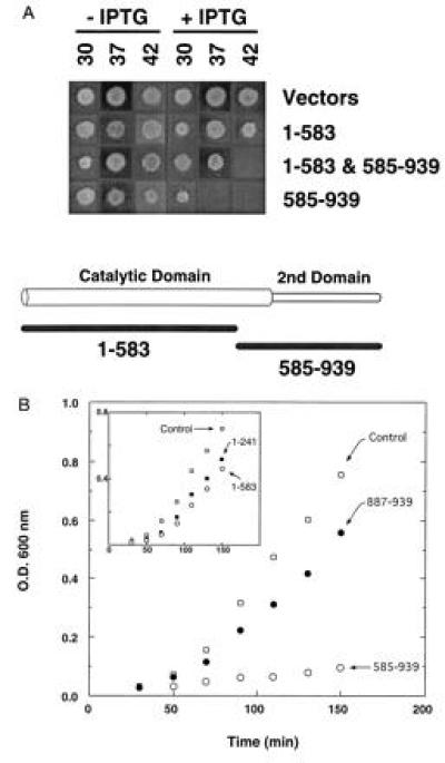
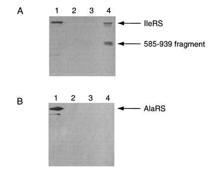
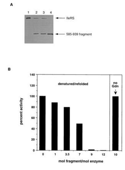
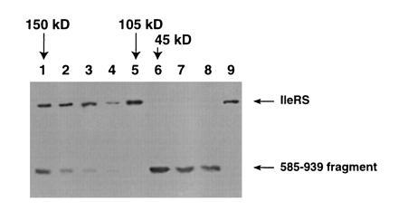
Dominant negative inhibition is most commonly seen when a mutant subunit of a multisubunit protein is coexpressed with the wild-type protein so that assembly of a functional oligomer is impaired. By analogy, it should be possible to interfere with the functional assembly of a monomeric enzyme by interfering with the folding pathway. Experiments in vitro by others suggested that fragments of a monomeric enzyme might be exploited for this purpose. We report here dominant negative inhibition of bacterial cell growth by expression of fragments of a tRNA synthetase. Inhibition is fragment-specific, as not all fragments cause inhibition. An inhibitory fragment characterized in more detail forms a specific complex with the intact enzyme in vivo, leading to enzyme inactivation. This fragment also associated stoichiometrically with the full-length enzyme in vitro after denaturation and refolding, and the resulting complex was catalytically inactive. Inhibition therefore appears to arise from an interruption in the folding pathway of the wild-type enzyme, thus suggesting a new strategy to design dominant negative inhibitors of monomeric enzymes.


Figures

Figure 1
Figure 1
Growth inhibition by fragment 585–939. (A) JM109 cells expressing isoleucyl-tRNA synthetase (IleRS) fragments 1–583 (from plasmid pKS171) or 585–939 (from pKS246) were grown on plates at 30°C, 37°C, and 42°C. (B) Growth inhibition in liquid culture by fragments 887–939 and 585–939, and by 1–241 and 1–583 (Inset).
Figure 2
Figure 2
Immunoprecipitation of enzyme complex. (A) Lane 1: lysate from JM109 cells. Lanes 2–4: anti-myc immunoprecipitation reactions (lane 2, JM109; lane 3, JM109 cells expressing IleRS; lane 4, JM109 cells expressing both IleRS and fragment 585–939). (B) The membranes were stripped of IleRS antibody and reprobed using anti-AlaRS antibody.
Figure 3
Figure 3
(A) Purification of fragment 585–939. Lysates from JM109/pKS246 cells were fractionated by ammonium sulfate precipitation and analyzed. Peak fractions from the chromatographic runs were analyzed by SDS/PAGE and anti-IleRS Western blotting. Lane 1, purified E. coli IleRS. Lane 2, Sephacryl S-300HR chromatography of the cell lysate. Lane 3, MonoQ chromatography of the cell lysate. Lane 4, MonoQ chromatography followed by Superose-12 gel filtration chromatography under denaturing conditions (6 M guanidine·HCl). Arrows indicate the positions of standards for IleRS and fragment 585–939 of IleRS. (B) Inhibition of IleRS activity by 585–939 fragment. Purified IleRS fragment 585–939 and full-length IleRS were denatured in 6 M guanidinium HCl and renatured. Samples were assayed for aminoacylation activity toward tRNAIle. Percentages of regained activity are expressed relative to a control with no fragment 585–939 (IleRS regains 100% of activity after unfolding and renaturation). In the right panel, a 10-fold excess of fragment 585–939 was incubated with IleRS without unfolding and refolding for 120 min.
Figure 4
Figure 4
Gel filtration chromatography of inhibited complex. Full-length IleRS was denatured in 6 M guanidinium HCl in the presence of a 12-fold excess of the 585–939 fragment. The sample was renatured, and aminoacylation activity was found to be abolished. The sample was applied to a Sephacryl S300 gel filtration column (120 ml bed volume) in 50 mM potassium phosphate (pH 7.5), 0.1 mM NaCl, 50 mM 2-mercaptoethanol. Peaks of absorbance at 280 nm were analyzed by Western blotting with anti-IleRS antibody. Lanes 1–7 show fractions eluting from the gel filtration column. Lane 8, fragment 585–939 of isoleucyl-tRNA synthetase; lane 9, pure isoleucyl-tRNA synthetase. The retention times of protein standards are indicated by arrows above the figure. Protein standards for the column were: alcohol dehydrogenase (150 kDa; elution volume = 33 ml), IleRS (105 kDa; elution volume = 39 ml), and ovalbumin (45 kDa; elution volume = 46 ml).

Similar articles

Cited by

References

    1. Herskowitz I. Nature (London) 1987;329:219–222. - PubMed
    1. Amaya E, Musci T J, Kirschner M W. Cell. 1991;66:257–270. - PubMed
    1. Verrall S, Hall Z W. Cell. 1992;68:23–31. - PubMed
    1. Kintner C. Cell. 1992;69:225–236. - PubMed
    1. Friedman A D, Triezenberg S J, McKnight S L. Nature (London) 1988;335:452–454. - PubMed

Publication types

MeSH terms

Substances

LinkOut - more resources