

Interactions of RecF protein with RecO, RecR, and single-stranded DNA binding proteins reveal roles for the RecF-RecO-RecR complex in DNA repair and recombination
- PMID: 8962075
- PMCID: PMC26156
- DOI: 10.1073/pnas.93.25.14468
Interactions of RecF protein with RecO, RecR, and single-stranded DNA binding proteins reveal roles for the RecF-RecO-RecR complex in DNA repair and recombination
Abstract
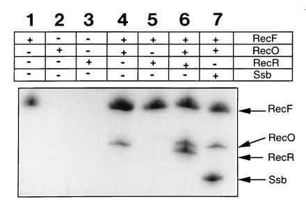
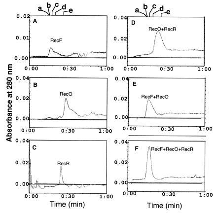
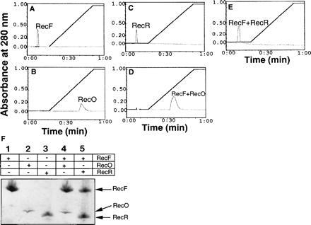
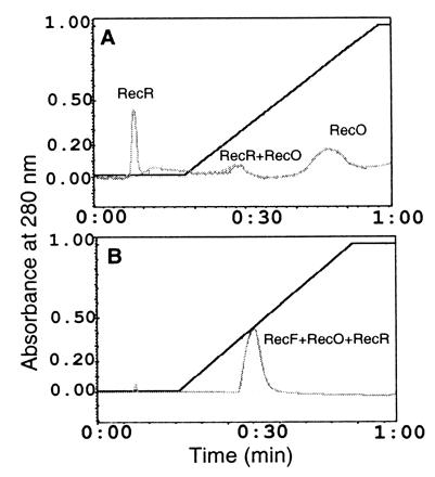
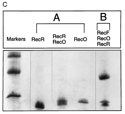
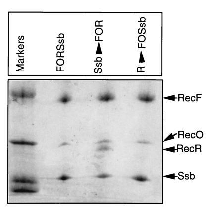
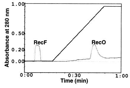
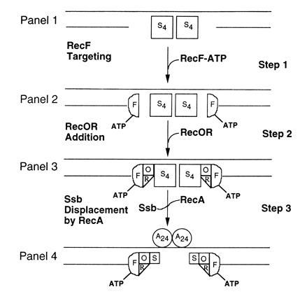
The products of the recF, recO, and recR genes are thought to interact and assist RecA in the utilization of single-stranded DNA precomplexed with single-stranded DNA binding protein (Ssb) during synapsis. Using immunoprecipitation, size-exclusion chromatography, and Ssb protein affinity chromatography in the absence of any nucleotide cofactors, we have obtained the following results: (i) RecF interacts with RecO, (ii) RecF interacts with RecR in the presence of RecO to form a complex consisting of RecF, RecO, and RecR (RecF-RecO-RecR); (iii) RecF interacts with Ssb protein in the presence of RecO. These data suggested that RecO mediates the interactions of RecF protein with RecR and with Ssb proteins. Incubation of RecF, RecO, RecR, and Ssb proteins resulted in the formation of RecF-RecO-Ssb complexes; i.e., RecR was excluded. Preincubation of RecF, RecO, and RecR proteins prior to addition of Ssb protein resulted in the formation of complexes consisting of RecF, RecO, RecR, and Ssb proteins. These data suggest that one role of RecF is to stabilize the interaction of RecR with RecO in the presence of Ssb protein. Finally, we found that interactions of RecF with RecO are lost in the presence of ATP. We discuss these results to explain how the RecF-RecO-RecR complex functions as an anti-Ssb factor.
Figures
Similar articles
-
Biochemical interaction of the Escherichia coli RecF, RecO, and RecR proteins with RecA protein and single-stranded DNA binding protein.Proc Natl Acad Sci U S A. 1993 May 1;90(9):3875-9. doi: 10.1073/pnas.90.9.3875. Proc Natl Acad Sci U S A. 1993. PMID: 8483906 Free PMC article.
-
Protein interactions in genetic recombination in Escherichia coli. Interactions involving RecO and RecR overcome the inhibition of RecA by single-stranded DNA-binding protein.J Biol Chem. 1994 Nov 25;269(47):30005-13. J Biol Chem. 1994. PMID: 7962001
-
The process of displacing the single-stranded DNA-binding protein from single-stranded DNA by RecO and RecR proteins.Nucleic Acids Res. 2008 Jan;36(1):94-109. doi: 10.1093/nar/gkm1004. Epub 2007 Nov 13. Nucleic Acids Res. 2008. PMID: 18000001 Free PMC article.
-
Regulation of bacterial RecA protein function.Crit Rev Biochem Mol Biol. 2007 Jan-Feb;42(1):41-63. doi: 10.1080/10409230701260258. Crit Rev Biochem Mol Biol. 2007. PMID: 17364684 Review.
-
Generation and Repair of Postreplication Gaps in Escherichia coli.Microbiol Mol Biol Rev. 2023 Jun 28;87(2):e0007822. doi: 10.1128/mmbr.00078-22. Epub 2023 May 22. Microbiol Mol Biol Rev. 2023. PMID: 37212693 Free PMC article. Review.
Cited by
-
The cell pole: the site of cross talk between the DNA uptake and genetic recombination machinery.Crit Rev Biochem Mol Biol. 2012 Nov-Dec;47(6):531-55. doi: 10.3109/10409238.2012.729562. Epub 2012 Oct 9. Crit Rev Biochem Mol Biol. 2012. PMID: 23046409 Free PMC article. Review.
-
Modulation of DNA repair and recombination by the bacteriophage lambda Orf function in Escherichia coli K-12.J Bacteriol. 2004 May;186(9):2699-707. doi: 10.1128/JB.186.9.2699-2707.2004. J Bacteriol. 2004. PMID: 15090511 Free PMC article.
-
Template-switching during replication fork repair in bacteria.DNA Repair (Amst). 2017 Aug;56:118-128. doi: 10.1016/j.dnarep.2017.06.014. Epub 2017 Jun 13. DNA Repair (Amst). 2017. PMID: 28641943 Free PMC article. Review.
-
Functional similarities between phage lambda Orf and Escherichia coli RecFOR in initiation of genetic exchange.Proc Natl Acad Sci U S A. 2005 Aug 9;102(32):11260-5. doi: 10.1073/pnas.0503399102. Epub 2005 Aug 2. Proc Natl Acad Sci U S A. 2005. PMID: 16076958 Free PMC article.
-
DNA repair and genome maintenance in Bacillus subtilis.Microbiol Mol Biol Rev. 2012 Sep;76(3):530-64. doi: 10.1128/MMBR.05020-11. Microbiol Mol Biol Rev. 2012. PMID: 22933559 Free PMC article. Review.
References
-
- Horii Z I, Clark A J. J Mol Biol. 1973;80:327–344. - PubMed
-
- Clark A J, Low K B. In: The Recombination of Genetic Material. Low K, editor. San Diego: Academic; 1988. pp. 155–215.
Publication types
MeSH terms
Substances
Grants and funding
LinkOut - more resources
Full Text Sources
Molecular Biology Databases
